
Page 141 - Read Online
P. 141
Page 8 of 12 Hallock. Plast Aesthet Res 2019;6:29 I http://dx.doi.org/10.20517/2347-9264.2019.029
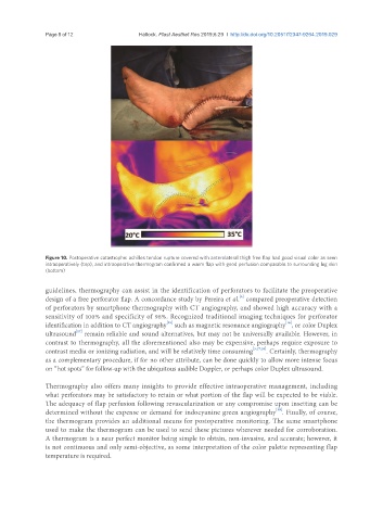
Figure 10. Postoperative catastrophe: achilles tendon rupture covered with anterolateral thigh free flap had good visual color as seen
intraoperatively (top), and intraoperative thermogram confirmed a warm flap with good perfusion comparable to surrounding leg skin
(bottom)
guidelines, thermography can assist in the identification of perforators to facilitate the preoperative
[6]
design of a free perforator flap. A concordance study by Pereira et al. compared preoperative detection
of perforators by smartphone thermography with CT angiography, and showed high accuracy with a
sensitivity of 100% and specificity of 98%. Recognized traditional imaging techniques for perforator
[16]
[15]
identification in addition to CT angiography such as magnetic resonance angiography , or color Duplex
[17]
ultrasound remain reliable and sound alternatives, but may not be universally available. However, in
contrast to thermography, all the aforementioned also may be expensive, perhaps require exposure to
contrast media or ionizing radiation, and will be relatively time consuming [3,17,18] . Certainly, thermography
as a complementary procedure, if for no other attribute, can be done quickly to allow more intense focus
on “hot spots” for follow-up with the ubiquitous audible Doppler, or perhaps color Duplex ultrasound.
Thermography also offers many insights to provide effective intraoperative management, including
what perforators may be satisfactory to retain or what portion of the flap will be expected to be viable.
The adequacy of flap perfusion following revascularization or any compromise upon insetting can be
[19]
determined without the expense or demand for indocyanine green angiography . Finally, of course,
the thermogram provides an additional means for postoperative monitoring. The same smartphone
used to make the thermogram can be used to send these pictures wherever needed for corroboration.
A thermogram is a near perfect monitor being simple to obtain, non-invasive, and accurate; however, it
is not continuous and only semi-objective, as some interpretation of the color palette representing flap
temperature is required.