
Page 142 - Read Online
P. 142
Hallock. Plast Aesthet Res 2019;6:29 I http://dx.doi.org/10.20517/2347-9264.2019.029 Page 9 of 12
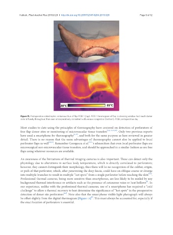
Figure 11. Postoperative catastrophe: violaceous hue of flap POD 1 (top). POD 1 thermogram of flap in dressing window had much darker
color diffusely throughout than seen intraoperatively consistent with venous congestion (bottom). POD: postoperative day
Most studies to date using the principles of thermography have centered on detection of perforators of
free flap donor sites or monitoring of microvascular tissue transfers [4,8,13,14,18] . Only two previous reports
have used a smartphone for thermography [6,11] , and both for the same purpose as here reviewed in greater
detail. There is no reason that the same advantages of thermography cannot also be applied to local
[22]
perforator flaps as well [20,21] . Remember Georgescu et al. ’s admonition that even local perforator flaps are
microsurgical non-microvascular tissue transfers, and should be approached in a similar fashion as are free
flaps using whatever resources are available.
An awareness of the limitations of thermal imaging cameras is also important. These can detect only the
physiology due to alterations in surface body temperature, which is directly correlated to perforators;
however, they cannot distinguish their morphology, thus there will be no recognition of the caliber, origin,
or path of that perforator, which, after penetrating the deep fascia, could have an oblique course or diverge
[5,8]
into multiple branches to result in multiple “hot spots” from a single perforator before reaching the skin .
Professional thermal cameras, being more sensitive than smartphones, are less likely to be misled by any
[8]
background thermal interference or artifacts such as the presence of cutaneous veins or heat hollows . In
our experience, unlike with the professional thermal cameras, use of a smartphone has required a “cold
challenge” to allow a thermal recovery to best determine the significance of “hot spots” in the preoperative
[3,5]
detection of donor site perforators . Note also that the smartphone visible light photograph will always
[7]
be offset slightly from the digital thermogram [Figure 13] . This must always be accounted for, especially if
the exact location of perforators is essential.