
Page 143 - Read Online
P. 143
Page 10 of 12 Hallock. Plast Aesthet Res 2019;6:29 I http://dx.doi.org/10.20517/2347-9264.2019.029
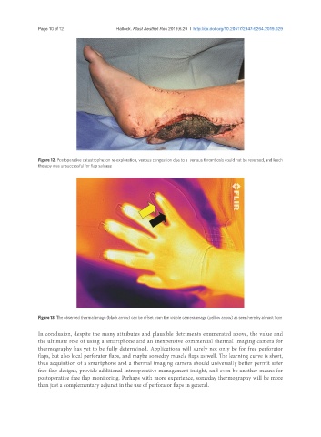
Figure 12. Postoperative catastrophe: on re-exploration, venous congestion due to a venous thrombosis could not be reversed, and leech
therapy was unsuccessful for flap salvage
Figure 13. The observed thermal image (black arrow) can be offset from the visible camera image (yellow arrow) as seen here by almost 1 cm
In conclusion, despite the many attributes and plausible detriments enumerated above, the value and
the ultimate role of using a smartphone and an inexpensive commercial thermal imaging camera for
thermography has yet to be fully determined. Applications will surely not only be for free perforator
flaps, but also local perforator flaps, and maybe someday muscle flaps as well. The learning curve is short,
thus acquisition of a smartphone and a thermal imaging camera should universally better permit safer
free flap designs, provide additional intraoperative management insight, and even be another means for
postoperative free flap monitoring. Perhaps with more experience, someday thermography will be more
than just a complementary adjunct in the use of perforator flaps in general.