
Page 139 - Read Online
P. 139
Page 6 of 12 Hallock. Plast Aesthet Res 2019;6:29 I http://dx.doi.org/10.20517/2347-9264.2019.029
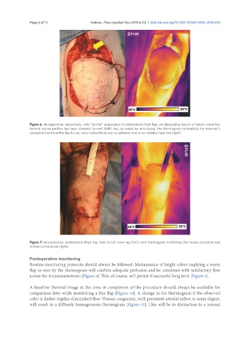
Figure 6. Intraoperative: subjectively, note “normal” appearance of anterolateral thigh flap, yet descending branch of lateral circumflex
femoral source pedicle has been clamped (arrow) (left), but, as would be anticipated, the thermogram contradicts the observer’s
assessment as the entire flap is cool, since indeed there was no perfusion and so no radiative heat loss (right)
Figure 7. Intraoperative: anterolateral thigh flap inset on left lower leg (left), with thermogram confirming that revascularization was
without compromise (right)
Postoperative monitoring
Routine monitoring protocols should always be followed. Maintenance of bright colors implying a warm
flap as seen by the thermogram will confirm adequate perfusion and be consistent with satisfactory flow
across the microanastomosis [Figure 8]. This, of course, will persist if successful long term [Figure 9].
A baseline thermal image at the time of completion of the procedure should always be available for
comparison later while monitoring a free flap [Figure 10]. A change in the thermogram if the observed
color is darker implies diminished flow. Venous congestion, with persistent arterial inflow to some degree,
will result in a diffusely homogeneous thermogram [Figure 11]. This will be in distinction to a normal