
Page 140 - Read Online
P. 140
Hallock. Plast Aesthet Res 2019;6:29 I http://dx.doi.org/10.20517/2347-9264.2019.029 Page 7 of 12
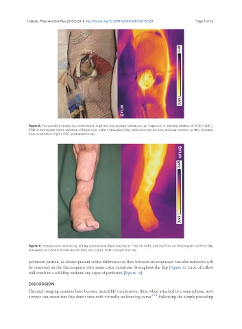
Figure 8. Postoperative monitoring: anterolateral thigh free flap appears satisfactory on inspection in dressing window on POD 2 (left ).
POD 2 thermogram shows variations of bright color pattern throughout flap, while dressings are dark, implying coolness, as they of course
have no perfusion (right). POD: postoperative day
Figure 9. Postoperative monitoring: left leg anterolateral thigh free flap at POD 40 (left), and the POD 40 thermogram confirms flap
adequately perfused and even warmer than toes (right). POD: postoperative day
perfusion pattern, as always present subtle differences in flow between encompassed vascular networks will
be observed on the thermogram with some color variations throughout the flap [Figure 8]. Lack of inflow
will result in a cold flap without any signs of perfusion [Figure 12].
DISCUSSION
Thermal imaging cameras have become incredibly inexpensive, thus, when attached to a smartphone, now
anyone can assess free flap donor sites with virtually no learning curve [6,11] . Following the simple preceding