
Page 136 - Read Online
P. 136
Hallock. Plast Aesthet Res 2019;6:29 I http://dx.doi.org/10.20517/2347-9264.2019.029 Page 3 of 12
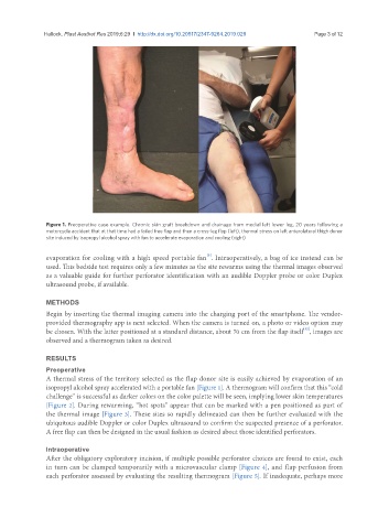
Figure 1. Preoperative case example. Chronic skin graft breakdown and drainage from medial left lower leg, 20 years following a
motorcycle accident that at that time had a failed free flap and then a cross-leg flap (left), thermal stress on left anterolateral thigh donor
site induced by isopropyl alcohol spray with fan to accelerate evaporation and cooling (right)
[5]
evaporation for cooling with a high speed portable fan . Intraoperatively, a bag of ice instead can be
used. This bedside test requires only a few minutes as the site rewarms using the thermal images observed
as a valuable guide for further perforator identification with an audible Doppler probe or color Duplex
ultrasound probe, if available.
METHODS
Begin by inserting the thermal imaging camera into the charging port of the smartphone. The vendor-
provided thermography app is next selected. When the camera is turned on, a photo or video option may
[12]
be chosen. With the latter positioned at a standard distance, about 70 cm from the flap itself , images are
observed and a thermogram taken as desired.
RESULTS
Preoperative
A thermal stress of the territory selected as the flap donor site is easily achieved by evaporation of an
isopropyl alcohol spray accelerated with a portable fan [Figure 1]. A thermogram will confirm that this “cold
challenge” is successful as darker colors on the color palette will be seen, implying lower skin temperatures
[Figure 2]. During rewarming, “hot spots” appear that can be marked with a pen positioned as part of
the thermal image [Figure 3]. These sites so rapidly delineated can then be further evaluated with the
ubiquitous audible Doppler or color Duplex ultrasound to confirm the suspected presence of a perforator.
A free flap can then be designed in the usual fashion as desired about those identified perforators.
Intraoperative
After the obligatory exploratory incision, if multiple possible perforator choices are found to exist, each
in turn can be clamped temporarily with a microvascular clamp [Figure 4], and flap perfusion from
each perforator assessed by evaluating the resulting thermogram [Figure 5]. If inadequate, perhaps more