
Page 56 - Read Online
P. 56
Baek et al. Plast Aesthet Res 2024;11:49 https://dx.doi.org/10.20517/2347-9264.2024.91 Page 7 of 11
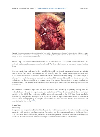
Figure 6. The gluteus maximus has been split along its fibers to follow along the course of two perforators indicated with black arrows.
Just inferior to the perforators and deep to the gluteus maximus is the piriformis (white asterisks). As the perforators are followed along
deeper, the plexus is encountered (white arrows).
After the flap has been successfully harvested, it can be further shaped on the back table while the donor site
is closed. Meticulous hemostasis should be achieved. The donor site is closed in layers over a closed suction
drain.
Microsurgery is then performed in the typical fashion with end-to-end venous anastomosis and arterial
anastomosis to the internal mammary vessels. We generally select the internal mammary vessels at the level
of the fourth rib or lower to minimize mismatch with the internal mammary artery. Inadequate length or
mismatch in vessel diameter may warrant vein grafts, which can be obtained from the saphenous vein,
cephalic vein, or the superficial inferior epigastric vein. Alternatively, the deep inferior epigastric pedicle can
be harvested via a low transverse abdominal incision as a composite vascular conduit to improve length and
vessel match.
For flap inset, a hammock-style inset has been described. This is done by suspending the flap onto the
pectoralis fascia obliquely in a superolateral and medial fashion [7,10] . As already described, due to the firmer
qualities of the SGAP flap, projection will be improved compared to the DIEP flap, but it also lacks
flexibility. By manipulating the flap with the tapered fat along the superior side of the breast reconstruction
and the firmer, more projecting fat along the caudal side of the reconstruction, the SGAP characteristics can
be optimized for the pocket [12,15,18] .
Sc-GAP flap
Positioning
The patient can be positioned in the lateral decubitus position as described above for simultaneous flap
harvesting with recipient vessel preparation. The patient can also be positioned prone. Rodriguez-Vegas
et al. found that the sc-GAP can be performed in the supine position, due to the more lateral and superior
[8]
location of the septocutaneous perforators compared to the musculocutaneous perforators .