
Page 55 - Read Online
P. 55
Page 6 of 11 Baek et al. Plast Aesthet Res 2024;11:49 https://dx.doi.org/10.20517/2347-9264.2024.91
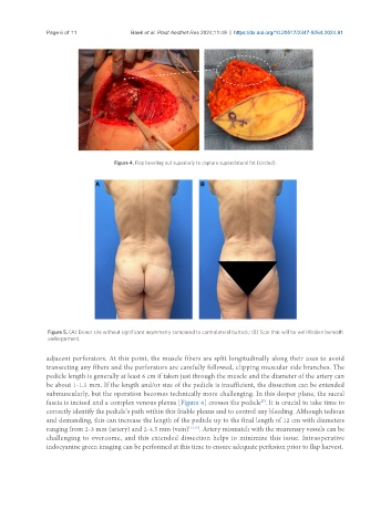
Figure 4. Flap beveling out superiorly to capture superolateral fat (circled).
Figure 5. (A) Donor site without significant asymmetry compared to contralateral buttock; (B) Scar that will be well-hidden beneath
undergarment.
adjacent perforators. At this point, the muscle fibers are split longitudinally along their axes to avoid
transecting any fibers and the perforators are carefully followed, clipping muscular side branches. The
pedicle length is generally at least 6 cm if taken just through the muscle and the diameter of the artery can
be about 1-1.5 mm. If the length and/or size of the pedicle is insufficient, the dissection can be extended
submuscularly, but the operation becomes technically more challenging. In this deeper plane, the sacral
fascia is incised and a complex venous plexus [Figure 6] crosses the pedicle . It is crucial to take time to
[7]
correctly identify the pedicle’s path within this friable plexus and to control any bleeding. Although tedious
and demanding, this can increase the length of the pedicle up to the final length of 12 cm with diameters
ranging from 2-3 mm (artery) and 2-4.5 mm (vein) [13,15] . Artery mismatch with the mammary vessels can be
challenging to overcome, and this extended dissection helps to minimize this issue. Intraoperative
indocyanine green imaging can be performed at this time to ensure adequate perfusion prior to flap harvest.