
Page 53 - Read Online
P. 53
Page 4 of 11 Baek et al. Plast Aesthet Res 2024;11:49 https://dx.doi.org/10.20517/2347-9264.2024.91
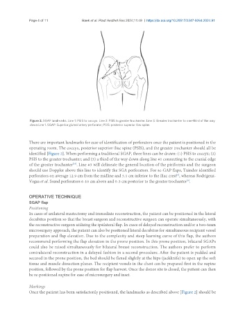
Figure 2. SGAP landmarks. Line 1: PSIS to coccyx. Line 2: PSIS to greater trochanter. Line 3: Greater trochanter to one-third of the way
down Line 1. SGAP: Superior gluteal artery perforator; PSIS: posterior superior iliac spine.
There are important landmarks for ease of identification of perforators once the patient is positioned in the
operating room. The coccyx, posterior superior iliac spine (PSIS), and the greater trochanter should all be
identified [Figure 2]. When performing a traditional SGAP, three lines can be drawn: (1) PSIS to coccyx; (2)
PSIS to the greater trochanter; and (3) a third of the way down along line #1 connecting to the cranial edge
of the greater trochanter . Line #3 will delineate the general location of the piriformis and the surgeon
[10]
should use Doppler above this line to identify the SGA perforators. For sc-GAP flaps, Tuinder identified
[9]
perforators on average 12.9 cm from the midline and 5.1 cm inferior to the iliac crest , whereas Rodrigeuz-
[8]
Vegas et al. found perforators 6-10 cm above and 0-3 cm posterior to the greater trochanter .
OPERATIVE TECHNIQUE
SGAP flap
Positioning
In cases of unilateral mastectomy and immediate reconstruction, the patient can be positioned in the lateral
decubitus position so that the breast surgeon and reconstructive surgeon can operate simultaneously, with
the reconstructive surgeon utilizing the ipsilateral flap. In cases of delayed reconstruction and/or a two-team
microsurgery approach, the patient can also be positioned lateral decubitus for simultaneous recipient vessel
preparation and flap elevation. Due to the complexity and steep learning curve of this flap, the authors
recommend performing the flap elevation in the prone position. In this prone position, bilateral SGAPs
could also be raised simultaneously for bilateral breast reconstruction. The authors prefer to perform
contralateral reconstruction in a delayed fashion in a second procedure. After the patient is padded and
secured in the prone position, the bed should be flexed slightly at the hips (jackknife) to open up the soft
tissue and muscle dissection planes. The recipient vessels in the chest can be prepared first in the supine
position, followed by the prone position for flap harvest. Once the donor site is closed, the patient can then
be re-positioned supine for ease of microsurgery and inset.
Markings
Once the patient has been satisfactorily positioned, the landmarks as described above [Figure 2] should be