
Page 54 - Read Online
P. 54
Baek et al. Plast Aesthet Res 2024;11:49 https://dx.doi.org/10.20517/2347-9264.2024.91 Page 5 of 11
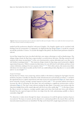
Figure 3. Elliptical skin design drawn around perforators identified with pencil Doppler. White arrow marks the lateral-most perforator.
Black lines indicate the planned beveling superiorly.
marked and the perforators identified with pencil Doppler. The Doppler signals can be correlated with
findings from the preoperative CT angiogram. An elliptical skin flap design [Figure 3] should be centered
around the perforator of choice. To increase the length of the pedicle, the lateral-most perforator should be
selected.
Several flap designs have been described with their own unique advantages as well as disadvantages. The
oblique design orients the flap from a superomedial to an inferolateral direction. This flap orientation will
[14]
optimize fatty tissue incorporation at the cost of postoperative contour deformity and a scar that is not
well-hidden in undergarments [13,14] . The transverse design orients the flap along a more horizontal to slightly
superolateral trajectory. While it contains less fatty tissue, the scar will often be well-hidden and there is less
contour deformity, making it the more popular choice [10,13,14,18] . With either pattern, the mastectomy defect
will dictate how much skin is needed, and the SGAP can comfortably provide a flap that is 8-12 cm in width
and 25-32 cm in length [6,15,18] .
Surgical technique
Because the fat is firmer, more projecting, and less pliable in the buttock, shaping the flap begins from the
moment of incision. Cranially, the dissection of the subcutaneous fat can be beveled out about 2-3 cm below
the superficial fascia to capture superolateral soft tissue excess and even lumbar fat [Figure 4]. Some authors
bevel inferiorly, thereby creating a mound tapered on both ends [13,14] . We minimize inferior beveling to
decrease the risk of hollowing out the donor site and resulting in a significant postoperative contour
deformity [Figure 5]. The flap is inset without rotating the skin island. Conversely, some authors take the
dissection straight down at the cranial edge and only bevel out at the caudal edge [12,15,18] . At the time of inset,
the flap is turned 180 degrees, creating a gentler upper pole slope that leads into lower pole fullness.
Blondeel has also reported improved donor site shape from the wide undermining caudally in what is
essentially a thigh lift .
[7]
The dissection is taken down through the deeper subcutaneous fat until the gluteus maximus fascia is
encountered. The fascia is incised, and the dissection is continued parallel to the muscle fibers until the
musculocutaneous perforators are identified. The flap can be based off a single larger perforator or several