
Page 52 - Read Online
P. 52
Baek et al. Plast Aesthet Res 2024;11:49 https://dx.doi.org/10.20517/2347-9264.2024.91 Page 3 of 11
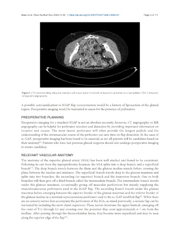
Figure 1. CTA demonstrating adequate available soft tissue donor in buttock compared to abdomen on a lean patient. CTA: Computed
tomography angiography.
A possible contraindication to SGAP flap reconstruction would be a history of liposuction of the gluteal
region. Preoperative imaging would be warranted to assess for the presence of perforators.
PREOPERATIVE PLANNING
Preoperative imaging for a standard SGAP is not an absolute necessity; however, CT angiography or MR
angiography can be helpful for perforator selection and dissection by providing important information on
location and course. The most lateral perforator will often provide the longest pedicle and the
understanding of the intramuscular course of the perforator can save time on flap dissection. In the cases of
sc-GAP, preoperative imaging has been found to be essential, as not all patients will be candidates based on
[8]
their anatomy . Patients who have had previous gluteal surgeries should also undergo preoperative imaging
to ensure candidacy.
RELEVANT VASCULAR ANATOMY
The anatomy of the superior gluteal artery (SGA) has been well studied and found to be consistent.
Following its exit from the suprapiriformis foramen, the SGA splits into a deep branch and a superficial
[16]
branch . The deep branch travels between the ilium and the gluteus medius muscle before crossing the
plane between the medius and minimus. The superficial branch travels deep to the gluteus maximus and
splits into two branches, the ascending (or superior) branch and the transverse branch. One or both
branches will then give off a third branch called the intermediate branch. The intermediate branch travels
under the gluteus maximus, occasionally giving off muscular perforators but mainly supplying the
musculocutaneous perforators used in the SGAP flap. The ascending branch travels under the gluteus
maximus before emerging between the superior border of the gluteus maximus and the inferior border of
[8]
the gluteus medius as a terminal septocutaneous perforator used in the sc-GAP modified flap . While there
are no sensory nerves that accompany the perforators of the SGA, as stated previously, a sensate flap can be
harvested by including the nervi clunii superioris. These nerves innervate the upper buttock, emerging off
the rami of T12 through L3 and crossing over the posterior iliac crest approximately 6-7 cm from the
midline. After passing through the thoracolumbar fascia, they become more superficial and may be seen
along the superior edge of the flap .
[17]