
Page 31 - Read Online
P. 31
Allam et al. Plast Aesthet Res 2024;11:19 https://dx.doi.org/10.20517/2347-9264.2024.21 Page 11 of 16
intra-muscular perforators and acquiring sufficient microsurgical experience to allow for seamless technical
execution. Additionally, the anastomosis of perforator pedicles to the internal mammary vessels within the
chest wall is especially difficult due to limited access and depth, which creates significant spatial challenges
for trainees. Currently, common microsurgery simulation models fail to adequately reflect these
challenges .
[60]
Consequently, there is a role for surgical education to pivot toward other effective and efficient teaching
strategies that can mirror the true complexity and subtleties of microsurgical techniques in breast
reconstruction.
Three-Dimensional (3D) printing
The application of 3D printing in surgical education transcends operative planning and offers a significant
educational advantage. Often, early-stage trainees struggle with the visuo-spatial skills required to interpret
CT angiography (CTA) imaging in the context of a patient's three-dimensional anatomy for perforator
dissection. In a pioneering effort, Mehta et al. utilized patient-specific 3D-printed models to replicate the
rectus abdominis muscle and the intricate intramuscular course of DIEA perforators. This model was
designed to assist trainees in evaluating patients’ three-dimensional surgical anatomy for effective
application in the operating room . Surgical residents were able to study the model preoperatively and
[15]
intraoperatively to help with perforator dissection. Anecdotal evidence indicated that these 3D models
enhanced clarity in dissection, offered superior visualization of the perforator’s course, and provided a
[15]
clearer understanding of perforator depth in comparison to traditional CTA imaging . This evidence is
supported by the success of previous 3D models used for education in skull base anatomy, complex orbital
anatomy, and even pathologic anatomy of craniofacial anomalies [62-64] .
As trainees progress in their understanding of perforator anatomy, they require consistent and realistic
methods for tactile practice outside the operating room. While the chicken thigh model is frequently used
given its similar vessel caliber to that of humans, it falls short in mimicking the actual clinical environment
and anatomical precision needed for successful technical execution . For instance, during chest wall
[60]
dissection and microvascular anastomosis, the depth at which the internal mammary vessels are located
poses significant training challenges in accessing and visualizing the anastomosis site, a complexity not
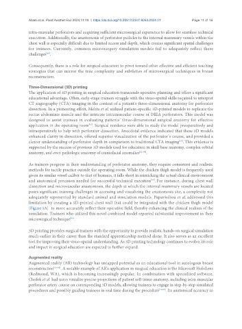
adequately represented by standard animal and simulation models. Papavisiliou et al. addressed this
limitation by creating a 3D-printed chest wall that could be integrated with the chicken thigh model
[Figure 5A] to more accurately reflect their operative field, thereby enhancing the clinical realism of the
simulation. Trainees who utilized this novel combined model reported substantial improvement in their
microsurgical technique .
[60]
3D printing provides surgical trainees with the opportunity to provide realistic hands-on surgical simulation
much earlier in their career than the standard apprenticeship method alone. It also serves as an excellent
tool for improving their visuo-spatial understanding. As 3D printing technology continues to evolve, its role
and impact in surgical education are expected to further expand.
Augmented reality
Augmented reality (AR) technology has untapped potential as an educational tool in autologous breast
reconstruction [65-68] . A notable example of AR's application in surgical education is the Microsoft HoloLens
(Redmond, WA), which is becoming increasingly popular. In combination with specialized software,
Cholok et al. had users visualize precise projections of patient soft tissue anatomy, including intra-muscular
perforator artery course on corresponding 3D models, allowing trainees to engage in step-by-step simulated
procedures and possibly guiding trainees in real time during the procedure [16,69] . Its anatomical accuracy in