
Page 28 - Read Online
P. 28
Page 8 of 16 Allam et al. Plast Aesthet Res 2024;11:19 https://dx.doi.org/10.20517/2347-9264.2024.21
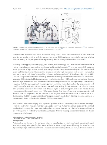
Figure 3. Intraoperative innovations. (A) MicroSure’s MUSA robotic platform (MicroSure, Eindhoven, Netherlands) [34] ; (B) Schematic
drawing of MMI Symani robotic platform (Medical Microinstruments, Pisa, Italy) [21] .
complications. Additionally, a proof-of-concept study created a real-time continuous in vitro perfusion
monitoring model with a high-frequency low-dose ICG regimen, potentially guiding clinical
[46]
decision-making in the perioperative setting after flap inset in autologous breast reconstruction .
With respect to hyperspectral imaging (HSI) system, this technology has advanced tissue visualization in
[47]
various surgical procedures, such as oncological and transplant surgeries . In broad terms, HSI captures a
broad spectrum of light waves, providing a comprehensive tissue assessment beyond the standard red,
green, and blue light waves. It can analyze tissue perfusion through parameters such as tissue oxygenation
patterns, near-infrared, tissue hemoglobin, and water perfusion markers . HSI offers an objective, reliable,
[48]
[49]
and user-independent method to assess flap perfusion in autologous breast reconstructions . Thiem et al.
translated HSI into the field of microsurgery, conducting the first HSI-based measurements on free and
pedicled flaps for head and neck reconstruction immediately after inset and postoperatively. Their findings
suggested that HSI can accurately measure perfusion viability immediately after flap anastomosis, with
specific cut-offs indicative of arterial/venous occlusion aiding decision-making on take-backs and
intraoperative revisions . Moreover, HSI detected signs of disturbed perfusion hours before clinical
[49]
symptoms manifested, and in one case, HSI analysis showed clear signs of emergent venous congestion 36 h
prior to clinical diagnosis . In the context of autologous breast reconstruction, Pruimboom et al.
[49]
demonstrated that HSI is superior in identifying postoperative mastectomy skin flap necrosis compared to
[25]
conventional clinical evaluation in unilateral DIEP reconstruction .
Both HSI and ICG-aided imaging have significantly advanced as reliable intraoperative tools for autologous
breast reconstructive surgery over the past decade. However, further research is necessary to establish
standardized protocols that could potentially reduce operation time and cost. Such advancements highlight
the evolving role of technology in enhancing surgical decision-making, traditionally reliant on clinical
experience and judgment.
POSTOPERATIVE INNOVATIONS
Background
Postoperative monitoring of flap perfusion is just as crucial a stage in autologous breast reconstruction as it
is intraoperatively. Complete flap loss is one of the most feared complications following this procedure, and
flap viability hinges on the integrity of the vascular anastomosis and patency. As such, early identification of