
Page 27 - Read Online
P. 27
Allam et al. Plast Aesthet Res 2024;11:19 https://dx.doi.org/10.20517/2347-9264.2024.21 Page 7 of 16
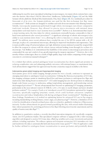
Addressing the need for more specialized technology in microsurgery, two main robotic systems have come
onto the market: MicroSure’s MUSA (MicroSure, Eindhoven, Netherlands) [Figure 3A] and the MMI
Symani robotic platform (Medical Microinstruments, Pisa, Italy) [Figure 3B]. In a landmark procedure by
Innocenti et al. in 2021, the Symani platform was used for the first-in-human free flap tissue
reconstruction . Both systems are designed to stabilize and scale down movements while filtering tremors.
[21]
Initially, microvascular anastomoses performed through robotic microsurgery were slower compared to
traditional methods. However, they allowed for precise micro-movements with minimal tissue
[33]
manipulation and made hard-to-reach anatomy more accessible . Barbon et al. demonstrated that despite
a steep learning curve, the time taken for robotic anastomosis eventually became comparable to that of
[35]
hand-sewn anastomosis after several procedures . A significant advantage of robotic microsurgery is the
ability to scale motions down from 7-20 x, allowing the robot to function at a slower, more controlled
speed . In addition, more recent advances have coupled the use of the MUSA system with a 4K 3D
[33]
exoscope, in place of a conventional microscope, allowing for improved visualization and ergonomics. This
is made possible using 3D polarized glasses and high-definition screens stationed around the surgical field
to allow the surgeon to operate with the robotic system without looking down through the oculars of a
microscope. Van Mulken et al. demonstrated the feasibility of this system in microsurgical procedures and
commended the ease and comfort of use, greatly improving the surgeon experience . However, due to the
[36]
novelty of these technologies, there is a dearth of high-quality, large-scale studies comparing robotic surgical
outcomes to conventional manual techniques .
[22]
It is evident that robotic-assisted autologous breast reconstruction has shown significant promise in
reducing complication rates and enhancing aesthetic outcomes, with minimal impact on anastomosis time.
Such advancements suggest that this approach may become a mainstay surgical modality in the future.
Indocyanine green-aided imaging and hyperspectral imaging
Indocyanine green (ICG)-aided imaging, though present for over a decade, continues to represent an
emerging innovation in autologous breast reconstruction. Utilizing the fluorescent properties of ICG dye,
this imaging technique assists in visualizing blood flow and tissue perfusion in autologous free flaps and
mastectomy skin during breast reconstruction . ICG-aided imaging has also shown promise in enhancing
[37]
lymph node biopsy accuracy in melanoma, aiding lymphedema management, and monitoring various
microsurgical procedures [38,39] . Recent advancements have focused on optimizing ICG imaging parameters,
particularly in the near-infrared window II (NIR-II, 1,000-1,700 nm), to enable deeper and more detailed
perfusion analysis . For example, Proulx et al. introduced a novel ICG formulation optimized for imaging
[40]
outcomes, specifically targeting lymphatic vessels, which could facilitate concurrent lymphatic
[26]
reconstruction and reduce lymphedema risk during autologous breast reconstruction . Building upon
these findings, Pruimboom et al. worked on optimizing factors for enhanced ICG use in flap surgery,
including standardizing working distance, sensor angle, and pharmacology . However, a limitation of ICG
[24]
is that the identification of poorly perfused areas relies on subjective, qualitative evaluation of fluorescence
intensity. Further advancements are geared toward quantifying the data, with several studies illustrating
time-related parameters for predicting perfusion in free flaps following microvascular anastomosis and the
quality of mastectomy skin flaps [41-43] .
In addition to quantitative analysis, a recently described innovation integrates AI, specifically computer
vision, into ICG perfusion studies. This specialized computer software is equipped with a video-tracking
algorithm and designed to analyze ICG tissue data, offsetting variables such as user movements and patient
artifacts, including respiration. The software captures and interprets various pixel ICG intensities over time
and transforms this data to automatically label and characterize tissue quality [44,45] . This AI application could
be further utilized in autologous breast reconstruction to assess perfusion and potentially detect