
Page 23 - Read Online
P. 23
Allam et al. Plast Aesthet Res 2024;11:19 https://dx.doi.org/10.20517/2347-9264.2024.21 Page 3 of 16
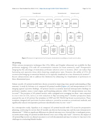
Figure 1. Framework of application of technological advancements in autologous breast reconstruction.
3D printing
While various preoperative techniques like CTA, MRA, and Doppler ultrasound are available for flap
[9]
perforator mapping, CTA with 3D reconstruction remains the most commonly used . Preoperative
imaging has the advantage of meticulous perforator selection prior to incision, which can reduce flap
harvesting and operative times while lowering complication rates. However, the utility of CTA 3D
[9]
reconstructed imaging is somewhat limited as it is typically visualized on a two-dimensional monitor .
Recent advancements aim to address this limitation by enhancing the visualization of perforators in
three-dimensional space.
Patient-specific 3D-printed models have shown potential in improving visuo-spatial understanding of flap
anatomy. A study by DeFazio et al. compared 3D-printed models [Figure 2A] and interpretations of CTA
imaging against operative findings. 3D-printed models accurately matched intraoperative findings in
perforator number, source vessel origin, and branching patterns, while CTA interpretations were less
[9]
accurate . The precision of 3D-printed models, with a marginal error averaging less than 1 mm compared
to over 8 mm in CTA, led to a 20 min reduction in flap harvesting time . Another prospective study
[9]
assessed the use of 3D-printed flap templates in DIEP and MS-TRAM flap breast reconstructions. These
templates, designed with perforator location indicators, proved as accurate as conventional methods, and
significantly reduced intraoperative perforator identification time by over 7 min .
[11]
In a retrospective study, Ogunleye et al. compared 3D-printed models with CTA scans for preoperative
planning, reinforcing the notion that 3D models can shorten harvesting time and decrease intraoperative
adjustments . While no significant differences in clinical outcomes were detected due to inadequate power,
[10]
[10]
reconstructions using 3D-printed models showed lower rates of flap take-back . However, the impact of
3D-printed flap models on clinical outcomes is not well-documented and may be subject to bias due to the
challenges in conducting blinded studies.