
Page 24 - Read Online
P. 24
Page 4 of 16 Allam et al. Plast Aesthet Res 2024;11:19 https://dx.doi.org/10.20517/2347-9264.2024.21
[9]
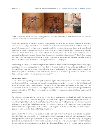
Figure 2. Preoperative planning. (A) Use of 3D model of epigastric perforators as a template for preoperative markings ; (B) Projection
of epigastric perforators onto patient abdominal wall [17] .
Despite their benefits, 3D-printed models for preoperative planning are not without limitations, including
cost, the use of a single material, and the exclusion of certain anatomical structures in earlier models [9-11] . A
proof-of-concept study by Jacobson et al. addressed this by combining voxel-based and mesh-based
modeling to create a more complex and accurate 3D-printed model of DIEP flaps, incorporating relevant
anatomical structures such as the rectus abdominis muscle, intra- and extra-muscular vessels, and a
geometric grid oriented around the umbilicus for anatomic localization of perforators . This hybrid model,
[14]
which accurately represented perforator locations compared to intraoperative findings, provided insights
that often differed from those based on interpretations of CTA imaging .
[14]
In summary, 3D-printed models offer significant added advantages over traditional preoperative imaging in
autologous breast reconstruction. However, their application is still in its nascent stages and has not yet
been widely adopted. Future research, including large-scale studies and cost analyses, is essential to establish
these techniques as a standard of care. Furthermore, the utility of anatomically complete 3D-printed DIEP
flaps in clinical practice remains an unexplored area [14,15] .
Augmented reality
AR is a novel and expanding technology that overlays digital information onto the real-world environment.
Applications of AR are becoming increasingly popular in surgery as they can improve the visualization of
anatomical structures by superimposing radiographic imaging onto the patient in real time. AR also
circumvents difficulties associated with 3D printing modalities and their need to be extrapolated to the
patient on the table. AR’s direct perioperative applicability by surgeons marks a significant technological
advancement.
To effectively integrate AR into clinical practice, its virtual projections must closely align with the patient's
anatomy. Cholok et al. validated the spatial congruence of AR projections with 3D-printed anatomical
models using the Microsoft HoloLens (Redmond, WA) technology . While they observed minor statistical
[16]
differences in translational displacement and axial rotation between the 3D models and virtual projections,
the overall scale of projection was accurate. This suggests that AR can reliably localize complex,
[16]
patient-specific anatomy during DIEP flap dissection .
Provided that AR projections can match spatial fidelity with patient anatomy, it is essential to determine
their feasibility and practicability in clinical practice. In a Danish case series involving five patients, AR was
used preoperatively to superimpose virtual projections over patients’ abdomen, identifying and mapping
perforators in comparison to ultrasound localization . Using the HoloLens, these projections were then
[12]
tagged, enalbing freedom of movement during the dissection. In four out of five cases, the point for which