
Page 102 - Read Online
P. 102
Page 170 Muroy et al. Neuroimmunol Neuroinflammation 2020;7:166-82 I http://dx.doi.org/10.20517/2347-8659.2020.16
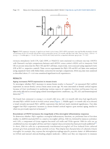
Figure 1. Phf15 expression increases in aged mouse frontal cortical areas. Phf15 mRNA expression was significantly elevated in frontal
cortical areas of old (~20-month-old; red bar) mice compared to young (~2.5-month-old; black bar) mice. Data are mean ± SEM (n = 4
young, n = 5 middle aged, n = 12 old). One-way ANOVA with Tukey’s post hoc comparisons between age groups: **P < 0.01
immune stimulation [with LPS, CpG-ODN, or Poly(I:C)] were analyzed via ordinary one-way ANOVA
with Dunnett’s multiple comparisons between each shRNA versus control shRNA within timepoint. Fold
OE or percent reduction for Phf15 OE and KO cell lines, respectively, were analyzed using unpaired t-tests
(OE or KO vs. respective control). Time course experiments for Phf15 OE and KO cell lines were analyzed
using unpaired t-tests with Holm-Sidak correction for multiple comparisons. RNA-seq data were analyzed
as described above. P < 0.05 was considered significant in all experiments.
RESULTS
Aging increases Phf15 expression in mouse brain
[21]
To investigate whether Phf15 increases in mouse brains similar to humans , we measured Phf15 mRNA
expression in mouse frontal cortical brain areas across age. We were interested in frontal cortical regions
because of their involvement in mediating various aspects of cognitive function and because they are
selectively affected in several aging-related neurodegenerative conditions, e.g., PD, AD, and frontotemporal
dementia [33,34] .
We found that compared to young (~2.5-month-old) mice, old (~20-month-old) mice had significantly
elevated Phf15 mRNA levels in frontal cortical areas [Figure 1]. Middle-aged (~14-month-old) mice showed
a trend towards increased Phf15 mRNA expression that did not reach statistical significance. Our data
suggest that Phf15 expression increases in mouse frontal cortical regions upon normal aging, similar to
[21]
what was previously reported in humans .
Knockdown of Phf15 increases the magnitude of the microglial inflammatory response
To determine whether Phf15 regulates microglial inflammatory function, we performed loss-of-function
studies via shRNA-mediated KD in a murine microglial cell line, SIM-A9, followed by immune activation
with LPS, a component of Gram-negative bacterial cell walls and TLR4 agonist. SIM-A9 cells are an
[35]
established murine microglial cell line and their original characterization has been previously published .
In short, SIM-A9 cells are a spontaneously transformed microglial cell line isolated from cultured
primary glia from postnatal murine cerebral cortices. They display key characteristics of cultured primary
microglia; for example, they express the microglia/macrophage-specific proteins cluster of differentiation
68 (CD68) and ionized calcium-binding adapter molecule 1 (IBA1) and they are responsive to immune