
Page 59 - Read Online
P. 59
Griffiths et al. Neuroimmunol Neuroinflammation 2020;7:51-67 I http://dx.doi.org/10.20517/2347-8659.2019.21 Page 55
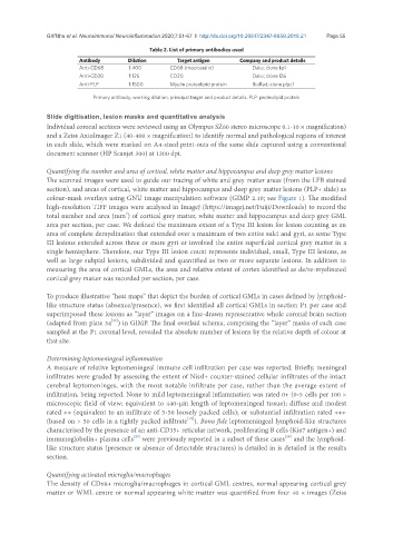
Table 2. List of primary antibodies used
Antibody Dilution Target antigen Company and product details
Anti-CD68 1:400 CD68 (macrosialin) Dako; clone kp1
Anti-CD20 1:125 CD20 Dako; clone l26
Anti-PLP 1:1500 Myelin proteolipid protein BioRad; clone plpc1
Primary antibody, working dilution, principal target and product details. PLP: proteolipid protein
Slide digitisation, lesion masks and quantitative analysis
Individual coronal sections were reviewed using an Olympus SZ60 stereo microscope 0.1-10 × magnification)
and a Zeiss AxioImager Z1 (40-400 × magnification) to identify normal and pathological regions of interest
in each slide, which were marked on A4-sized print-outs of the same slide captured using a conventional
document scanner (HP Scanjet 300) at 1200 dpi.
Quantifying the number and area of cortical, white matter and hippocampus and deep grey matter lesions
The scanned images were used to guide our tracing of white and grey matter areas (from the LFB stained
section), and areas of cortical, white matter and hippocampus and deep grey matter lesions (PLP+ slide) as
colour-mask overlays using GNU image manipulation software (GIMP 2.10; see Figure 1). The modified
high-resolution TIFF images were analysed in ImageJ (https://imagej.net/Fuiji/Downloads) to record the
2
total number and area (mm ) of cortical grey matter, white matter and hippocampus and deep grey GML
area per section, per case. We defined the maximum extent of a Type III lesion for lesion counting as an
area of complete demyelination that extended over a maximum of two entire sulci and gyri, as some Type
III lesions extended across three or more gyri or involved the entire superficial cortical grey matter in a
single hemisphere. Therefore, our Type III lesion count represents individual, small, Type III lesions, as
well as large subpial lesions, subdivided and quantified as two or more separate lesions. In addition to
measuring the area of cortical GMLs, the area and relative extent of cortex identified as de/re-myelinated
cortical grey matter was recorded per section, per case.
To produce illustrative “heat maps” that depict the burden of cortical GMLs in cases defined by lymphoid-
like structure status (absence/presence), we first identified all cortical GMLs in section P1 per case and
superimposed these lesions as “layer” images on a line-drawn representative whole coronal brain section
[22]
(adapted from plate 34 ) in GIMP. The final overlaid schema, comprising the “layer” masks of each case
sampled at the P1 coronal level, revealed the absolute number of lesions by the relative depth of colour at
that site.
Determining leptomeningeal inflammation
A measure of relative leptomeningeal immune cell infiltration per case was reported. Briefly, meningeal
infiltrates were graded by assessing the extent of Nissl+ counter-stained cellular infiltrates of the intact
cerebral leptomeninges, with the most notable infiltrate per case, rather than the average extent of
infiltration, being reported. None to mild leptomeningeal inflammation was rated 0+ (0-5 cells per 100 ×
microscopic field of view; equivalent to 440-µm length of leptomeningeal tissue); diffuse and modest
rated ++ (equivalent to an infiltrate of 5-50 loosely packed cells); or substantial infiltration rated +++
[18]
(based on > 50 cells in a tightly packed infiltrate ). Bona fide leptomeningeal lymphoid-like structures
characterised by the presence of an anti-CD35+ reticular network, proliferating B cells (Ki67 antigen+) and
immunoglobulin+ plasma cells were previously reported in a subset of these cases and the lymphoid-
[23]
[18]
like structure status (presence or absence of detectable structures) is detailed in is detailed in the results
section.
Quantifying activated microglia/macrophages
The density of CD68+ microglia/macrophages in cortical GML centres, normal appearing cortical grey
matter or WML centre or normal appearing white matter was quantified from four 40 × images (Zeiss