
Page 58 - Read Online
P. 58
Page 54 Griffiths et al. Neuroimmunol Neuroinflammation 2020;7:51-67 I http://dx.doi.org/10.20517/2347-8659.2019.21
A
B
C
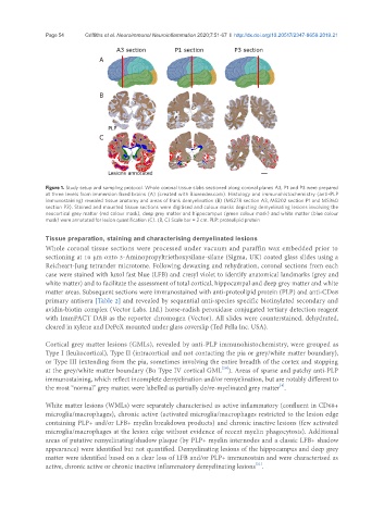
Figure 1. Study setup and sampling protocol. Whole coronal tissue slabs sectioned along coronal planes A3, P1 and P3 were prepared
at three levels from immersion fixed brains (A) (created with Biorender.com). Histology and immunohistochemistry (anti-PLP
immunostaining) revealed tissue anatomy and areas of frank demyelination (B) (MS278 section A3, MS202 section P1 and MS360
section P3). Stained and mounted tissue sections were digitised and colour masks depicting demyelinating lesions involving the
neocortical grey matter (red colour mask), deep grey matter and hippocampus (green colour mask) and white matter (blue colour
mask) were annotated for lesion quantification (C). (B, C) Scale bar = 2 cm. PLP: proteolipid protein
Tissue preparation, staining and characterising demyelinated lesions
Whole coronal tissue sections were processed under vacuum and paraffin wax embedded prior to
sectioning at 10 µm onto 3-Aminopropyltriethoxysilane-silane (Sigma, UK) coated glass slides using a
Reicheart-Jung tetrander microtome. Following dewaxing and rehydration, coronal sections from each
case were stained with luxol fast blue (LFB) and cresyl violet to identify anatomical landmarks (grey and
white matter) and to facilitate the assessment of total cortical, hippocampal and deep grey matter and white
matter areas. Subsequent sections were immunostained with anti-proteolipid protein (PLP) and anti-CD68
primary antisera [Table 2] and revealed by sequential anti-species specific biotinylated secondary and
avidin-biotin complex (Vector Labs. Ltd.) horse-radish peroxidase conjugated tertiary detection reagent
with ImmPACT DAB as the reporter chromogen (Vector). All slides were counterstained, dehydrated,
cleared in xylene and DePeX mounted under glass coverslip (Ted Pella Inc. USA).
Cortical grey matter lesions (GMLs), revealed by anti-PLP immunohistochemistry, were grouped as
Type I (leukocortical), Type II (intracortical and not contacting the pia or grey/white matter boundary),
or Type III (extending from the pia, sometimes involving the entire breadth of the cortex and stopping
[20]
at the grey/white matter boundary (Bo Type IV cortical GML ). Areas of sparse and patchy anti-PLP
immunostaining, which reflect incomplete demyelination and/or remyelination, but are notably different to
[4]
the most “normal” grey matter, were labelled as partially de/re-myelinated grey matter .
White matter lesions (WMLs) were separately characterised as active inflammatory (confluent in CD68+
microglia/macrophages), chronic active (activated microglia/macrophages restricted to the lesion edge
containing PLP+ and/or LFB+ myelin breakdown products) and chronic inactive lesions (few activated
microglia/macrophages at the lesion edge without evidence of recent myelin phagocytosis). Additional
areas of putative remyelinating/shadow plaque (by PLP+ myelin internodes and a classic LFB+ shadow
appearance) were identified but not quantified. Demyelinating lesions of the hippocampus and deep grey
matter were identified based on a clear loss of LFB and/or PLP+ immunostain and were characterised as
active, chronic active or chronic inactive inflammatory demyelinating lesions .
[21]