
Page 57 - Read Online
P. 57
Griffiths et al. Neuroimmunol Neuroinflammation 2020;7:51-67 I http://dx.doi.org/10.20517/2347-8659.2019.21 Page 53
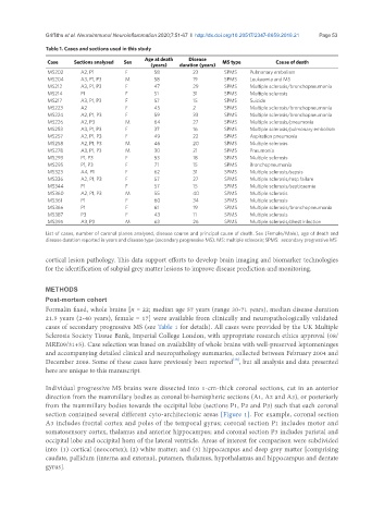
Table 1. Cases and sections used in this study
Age at death Disease
Case Sections analysed Sex MS type Cause of death
(years) duration (years)
MS202 A2, P1 F 58 23 SPMS Pulmonary embolism
MS204 A3, P1, P3 M 58 19 SPMS Leukaemia and MS
MS212 A3, P1, P3 F 47 29 SPMS Multiple sclerosis/bronchopneumonia
MS214 P1 F 51 31 SPMS Multiple sclerosis
MS217 A3, P1, P3 F 57 15 SPMS Suicide
MS223 A2 F 45 2 SPMS Multiple sclerosis/bronchopneumonia
MS224 A2, P1, P3 F 59 33 SPMS Multiple sclerosis/bronchopneumonia
MS226 A2, P3 M 64 27 SPMS Multiple sclerosis/pneumonia
MS253 A3, P1, P3 F 37 16 SPMS Multiple sclerosis/pulmonary embolism
MS257 A2, P1, P3 F 49 22 SPMS Aspiration pneumonia
MS258 A2, P1, P3 M 46 20 SPMS Multiple sclerosis
MS278 A3, P1, P3 M 30 21 SPMS Pneumonia
MS293 P1, P3 F 53 18 SPMS Multiple sclerosis
MS295 P1, P3 F 71 15 SPMS Bronchopneumonia
MS323 A4, P1 F 62 31 SPMS Multiple sclerosis/sepsis
MS336 A2, P1, P3 F 57 27 SPMS Multiple sclerosis/resp failure
MS344 P1 F 57 15 SPMS Multiple sclerosis/septicaemia
MS360 A2, P1, P3 M 55 40 SPMS Multiple sclerosis
MS361 P1 F 60 34 SPMS Multiple sclerosis
MS366 P1 F 61 19 SPMS Multiple sclerosis/bronchopneumonia
MS387 P3 F 43 11 SPMS Multiple sclerosis
MS395 A3, P3 M 63 26 SPMS Multiple sclerosis/chest infection
List of cases, number of coronal planes analysed, disease course and principal cause of death. Sex (Female/Male), age of death and
disease duration reported in years and disease type (secondary progressive MS). MS: multiple sclerosis; SPMS: secondary progressive MS
cortical lesion pathology. This data support efforts to develop brain imaging and biomarker technologies
for the identification of subpial grey matter lesions to improve disease prediction and monitoring.
METHODS
Post-mortem cohort
Formalin fixed, whole brains [n = 22; median age 57 years (range 30-71 years), median disease duration
21.5 years (2-40 years), female = 17] were available from clinically and neuropathologically validated
cases of secondary progressive MS (see Table 1 for details). All cases were provided by the UK Multiple
Sclerosis Society Tissue Bank, Imperial College London, with appropriate research ethics approval (08/
MRE09/31+5). Case selection was based on availability of whole brains with well-preserved leptomeninges
and accompanying detailed clinical and neuropathology summaries, collected between February 2004 and
December 2008. Some of these cases have previously been reported , but all analysis and data presented
[18]
here are unique to this manuscript.
Individual progressive MS brains were dissected into 1-cm-thick coronal sections, cut in an anterior
direction from the mammillary bodies as coronal bi-hemispheric sections (A1, A2 and A3), or posteriorly
from the mammillary bodies towards the occipital lobe (sections P1, P2 and P3) such that each coronal
section contained several different cyto-architectonic areas [Figure 1]. For example, coronal section
A3 includes frontal cortex and poles of the temporal gyrus; coronal section P1 includes motor and
somatosensory cortex, thalamus and anterior hippocampus; and coronal section P3 includes parietal and
occipital lobe and occipital horn of the lateral ventricle. Areas of interest for comparison were subdivided
into: (1) cortical (neocortex); (2) white matter; and (3) hippocampus and deep grey matter [comprising
caudate, pallidum (interna and externa), putamen, thalamus, hypothalamus and hippocampus and dentate
gyrus].