
Page 143 - Read Online
P. 143
Page 8 of 10 Alam et al. Neuroimmunol Neuroinflammation 2018;5:21 I http://dx.doi.org/10.20517/2347-8659.2017.64
A B
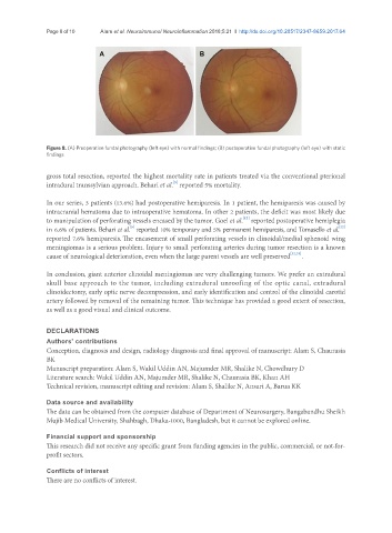
Figure 8. (A) Preoperative fundal photography (left eye) with normal findings; (B) postoperative fundal photography (left eye) with static
findings
gross total resection, reported the highest mortality rate in patients treated via the conventional pterional
[5]
intradural transsylvian approach. Behari et al. reported 5% mortality.
In our series, 3 patients (13.6%) had postoperative hemiparesis. In 1 patient, the hemiparesis was caused by
intracranial hematoma due to intraoperative hematoma. In other 2 patients, the deficit was most likely due
[12]
to manipulation of perforating vessels encased by the tumor. Goel et al. reported postoperative hemiplegia
[5]
[22]
in 6.6% of patients. Behari et al. reported 10% temporary and 5% permanent hemiparesis, and Tomasello et al.
reported 7.6% hemiparesis. The encasement of small perforating vessels in clinoidal/medial sphenoid wing
meningiomas is a serious problem. Injury to small perforating arteries during tumor resection is a known
cause of neurological deterioration, even when the large parent vessels are well preserved [32,33] .
In conclusion, giant anterior clinoidal meningiomas are very challenging tumors. We prefer an extradural
skull base approach to the tumor, including extradural unroofing of the optic canal, extradural
clinoidectomy, early optic nerve decompression, and early identification and control of the clinoidal carotid
artery followed by removal of the remaining tumor. This technique has provided a good extent of resection,
as well as a good visual and clinical outcome.
DECLARATIONS
Authors’ contributions
Conception, diagnosis and design, radiology diagnosis and final approval of manuscript: Alam S, Chaurasia
BK
Manuscript preparation: Alam S, Wakil Uddin AN, Majumder MR, Shalike N, Chowdhury D
Literature search: Wakil Uddin AN, Majumder MR, Shalike N, Chaurasia BK, Khan AH
Technical revision, manuscript editing and revision: Alam S, Shalike N, Ansari A, Barua KK
Data source and availability
The data can be obtained from the computer database of Department of Neurosurgery, Bangabandhu Sheikh
Mujib Medical University, Shahbagh, Dhaka-1000, Bangladesh, but it cannot be explored online.
Financial support and sponsorship
This research did not receive any specific grant from funding agencies in the public, commercial, or not-for-
profit sectors.
Conflicts of interest
There are no conflicts of interest.