
Page 138 - Read Online
P. 138
Alam et al. Neuroimmunol Neuroinflammation 2018;5:21 I http://dx.doi.org/10.20517/2347-8659.2017.64 Page 3 of 10
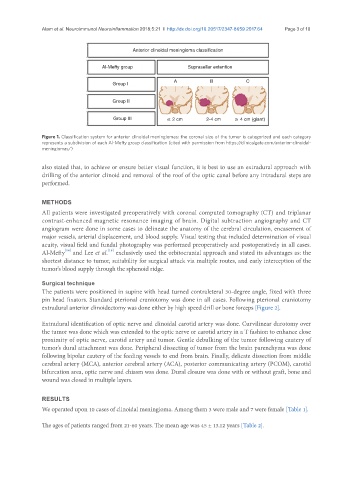
Anterior clinoidal meningioma classification
Al-Mefty group Suprasellar extention
A B C
Group I
Group II
Group III ≤ 2 cm 2-4 cm ≥ 4 cm (giant)
Figure 1. Classification system for anterior clinoidal meningiomas: the coronal size of the tumor is categorized and each category
represents a subdivision of each Al-Mefty group classification (cited with permission from https://clinicalgate.com/anterior-clinoidal-
meningiomas/)
also stated that, to achieve or ensure better visual function, it is best to use an extradural approach with
drilling of the anterior clinoid and removal of the roof of the optic canal before any intradural steps are
performed.
METHODS
All patients were investigated preoperatively with coronal computed tomography (CT) and triplanar
contrast-enhanced magnetic resonance imaging of brain. Digital subtraction angiography and CT
angiogram were done in some cases to delineate the anatomy of the cerebral circulation, encasement of
major vessels, arterial displacement, and blood supply. Visual testing that included determination of visual
acuity, visual field and fundal photography was performed preoperatively and postoperatively in all cases.
[18]
[14]
Al-Mefty and Lee et al. exclusively used the orbitocranial approach and stated its advantages as: the
shortest distance to tumor, suitability for surgical attack via multiple routes, and early interception of the
tumor’s blood supply through the sphenoid ridge.
Surgical technique
The patients were positioned in supine with head turned contraleteral 30-degree angle, fixed with three
pin head fixators. Standard pterional craniotomy was done in all cases. Following pterional craniotomy
extradural anterior clinoidectomy was done either by high speed drill or bone forceps [Figure 2].
Extradural identification of optic nerve and clinoidal carotid artery was done. Curvilinear durotomy over
the tumor was done which was extended to the optic nerve or carotid artery in a T fashion to enhance close
proximity of optic nerve, carotid artery and tumor. Gentle debulking of the tumor following cautery of
tumor’s dural attachment was done. Peripheral dissecting of tumor from the brain parenchyma was done
following bipolar cautery of the feeding vessels to end from brain. Finally, delicate dissection from middle
cerebral artery (MCA), anterior cerebral artery (ACA), posterior communicating artery (PCOM), carotid
bifurcation area, optic nerve and chiasm was done. Dural closure was done with or without graft, bone and
wound was closed in multiple layers.
RESULTS
We operated upon 10 cases of clinoidal meningioma. Among them 3 were male and 7 were female [Table 1].
The ages of patients ranged from 21-60 years. The mean age was 45 ± 13.12 years [Table 2].