
Page 142 - Read Online
P. 142
Alam et al. Neuroimmunol Neuroinflammation 2018;5:21 I http://dx.doi.org/10.20517/2347-8659.2017.64 Page 7 of 10
A B
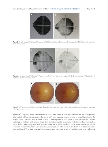
Figure 5. Humphrey visual field test. (A) Preoperative VF (right eye) with total blindness; (B) postoperative VF (right eye) with significant
visual improvement
A B
Figure 6. Humphrey visual field test. (A) Preoperative VF (left eye) with upper temporal field; (B) postoperative VF (left eye) with
significant visual improvement defect
A B
Figure 7. (A) Preoperative fundal photography (right eye) with primary optic atrophy; (B) postoperative fundal photography (right eye)
with slight improvement
[30]
Benjamin reported visual improvement in 73%, stable vision in 20%, and deterioration in 7% of patients
[31]
who had visual loss before surgery. Pamir et al. also reported improvements in visual function in the
majority of 43 patients with anterior clinoidal meningiomas with a mean tumor diameter of 3.35 cm,
including 16 patients with tumors larger than 4 cm in diameter. Among 26 patients who had preoperative
visual deficits, 84.6% improved and 15.4% remained stable. The Russell and Pamir teams used an intradural
approach. In our series, there were no deaths, in comparison with 5%-15.4% mortality in some recent series.
[22]
Tomasello et al. , whose patients had a mean tumor diameter of 5.7 cm and of whom 77% underwent