
Page 141 - Read Online
P. 141
Page 6 of 10 Alam et al. Neuroimmunol Neuroinflammation 2018;5:21 I http://dx.doi.org/10.20517/2347-8659.2017.64
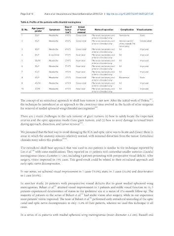
Table 6. Profile of the patients with clinoidal meningioma
Size of Extent
Sl. No. Age (years)/ Symptoms tumor of tumor Name of operation Complication Visual outcome
gender (cm ) removal
3
1 50/F Headache 6*6*6 Gross total Pterional craniotomy and Hematoma Static
anterior clinoidectomy
2 45/F Headache 8*6*5 Gross total Pterional craniotomy and Internal carotid Deteriorated
anterior clinoidectomy artery injured, 3rd
nerve palsy
3 50/F Headache 6*6*5 Gross total Pterional craniotomy and Nil Improved
anterior clinoidectomy
4 25/F Lt eye blind 6*5*5 Near total Pterional craniotomy and Nil Improved
anterior clinoidectomy
5 58/M Headache 5*4*5 Near total Pterional craniotomy and Nil Improved
anterior clinoidectomy
6 36/F Headache 5*4*5 Near total Pterional craniotomy and Nil Improved
anterior clinoidectomy
7 42/F Headache 4*5*4 Near total Pterional craniotomy and Nil Improved
anterior clinoidectomy
8 45/F Headache 4*5*5 Gross total Pterional craniotomy and Recurrence Static
anterior clinoidectomy
9 45/M Headache 6*5*4 Gross total Pterional craniotomy and Nil Improved
anterior clinoidectomy
10 47/M Headache 6*5*4 Near total Pterional craniotomy and Nil Improved
anterior clinoidectomy
[11]
The concept of an extradural approach to skull base tumors is not new. After the initial work of Dolenc ,
the technique he introduced as an approach to the cavernous sinus evolved in the hands of other surgeons
[26]
for removal of medial sphenoid wing/clinoidal meningiomas .
There are 2 main challenges in the safe removal of giant tumors: (1) how to safely locate the important
arteries and the optic apparatus inside these giant tumors, and (2) how to avoid damage to tensed brain
[27]
during approach, dissection, and tumor removal .
We presumed that the best way to avoid damaging the ICA and optic nerve was to locate and dissect them in
areas in which the anatomy remains relatively normal, with minimal distortion from the tumor. Extradural
clinoidectomy solves this problem [28,29] .
The extradural skull base approach that was used in our patients is similar to the technique reported by
[18]
Lee et al. with some modifications. They reported on 15 patients with somewhat smaller anterior clinoidal
meningiomas (mean diameter 3.7 cm), including 8 patients presenting with preoperative visual deficits. After
surgery, vision improved in 75% cases. This good result could be related to their extradural approach and
early optic nerve decompression.
In our series, we achieved visual improvement in 7 cases (70.0%), static in 2 cases (20.0%) and deterioration
in 1 case (10.0%).
In another study, 20 patients with preoperative visual deficits due to giant medial sphenoid wing
[5]
meningiomas, Behari et al. attained visual improvement in 3 patients and stable visual function in 11; 5
patients experienced deterioration of vision in the ipsilateral eye at a mean of 17.6-month follow-up. The
[5]
majority of patients in the series of Behari et al. had stable vision after surgery, while in our experience
[5]
most patients’ vision improved. The team of Behari et al. performed early extradural unroofing of the optic
canal and optic nerve decompression in only 15.8% of their patients, whereas we used this technique in all
cases.
In a series of 35 patients with medial sphenoid wing meningiomas (mean diameter 4.5 cm), Russell and