
Page 139 - Read Online
P. 139
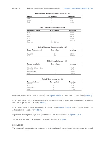
Page 4 of 10                  Alam et al. Neuroimmunol Neuroinflammation 2018;5:21  I  http://dx.doi.org/10.20517/2347-8659.2017.64


                                         Table 1. The distribution of patients by gender (n = 10)
                                Gender                  No. of patients        Percentage
                                Male                       3                    30.0%
                                Female                     7                    70.0%
                                Total                      10                   100.0%


                                              Table 2. The age of the patients (n = 10)
                                Age group (in years)      No. of patients      Percentage
                                21-30                      1                     10.0%
                                31-40                      1                     10.0%
                                41-50                      7                     70.0%
                                51-60                      1                     10.0%
                                Total                      10                    100.0%
                                Mean age                   45 ± 13.12 years


                                            Table 3. The extent of tumor removal (n = 10)
                                Extent of tumor removal    No. of patient       Percentage
                                Gross total                    5                 50.0%
                                Near total                     5                 50.0%
                                Total                          10                100.0%

                                                      Table 4. Complications (n = 10)

                                Name of complication       No. of patients      Percentage
                                Hematoma                       1                 10.0%
                                Internal carotid artery injured  1               10.0%
                                Recurrence                     1                 10.0%
                                No complication                7                 70.0%
                                Total                          10                100.0%

                                                 Table 5. Visual outcome (n = 10)
                                Functional outcome        No. of patients      Percentage
                                Improved                     7                   70.0%
                                Static                       2                   20.0%
                                Deteriorated                 1                   10.0%
                                Total                        10                  100.0%

               Gross total removal was achieved in 5 (50.0%) cases [Figures 3 and 4], and near total in 5 cases (50.0%) [Table 3].


               In our study most of the patients had limited complications. Only one patient had complicated by hematoma
               and another, patient had ICA injury [Table 4].


               In our series, we found visual improvement in 7 cases (70.0%) [Figures 5 and 6], static in 2 cases (20.0%), and
               deterioration in 1 case (10.0%) [Table 5].


               Papilledema also improved significantly after removal of tumors as shown in Figures 7 and 8.

               The profile of the patients with clinoidal meningioma is shown in Table 6.


               DISCUSSION
               The traditional approach for the resection of anterior clinoidal meningiomas is the pterional intradural