
Page 131 - Read Online
P. 131
Page 2 of 6 Verma et al. Neuroimmunol Neuroinflammation 2018;5:20 I http://dx.doi.org/10.20517/2347-8659.2017.65
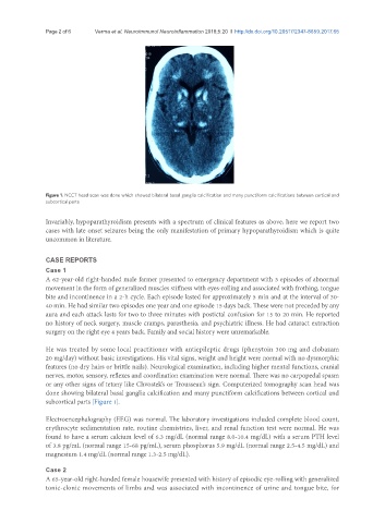
Figure 1. NCCT head scan was done which showed bilateral basal ganglia calcification and many punctiform calcifications between cortical and
subcortical parts
Invariably, hypoparathyroidism presents with a spectrum of clinical features as above, here we report two
cases with late onset seizures being the only manifestation of primary hypoparathyroidism which is quite
uncommon in literature.
CASE REPORTS
Case 1
A 62-year-old right-handed male farmer presented to emergency department with 3 episodes of abnormal
movement in the form of generalized muscles stiffness with eyes-rolling and associated with frothing, tongue
bite and incontinence in a 2-h cycle. Each episode lasted for approximately 5 min and at the interval of 30-
40 min. He had similar two episodes one year and one episode 15 days back. These were not preceded by any
aura and each attack lasts for two to three minutes with postictal confusion for 15 to 20 min. He reported
no history of neck surgery, muscle cramps, parasthesia, and psychiatric illness. He had cataract extraction
surgery on the right eye 4 years back. Family and social history were unremarkable.
He was treated by some local practitioner with antiepileptic drugs (phenytoin 300 mg and clobazam
20 mg/day) without basic investigations. His vital signs, weight and height were normal with no dysmorphic
features (no dry hairs or brittle nails). Neurological examination, including higher mental functions, cranial
nerves, motor, sensory, reflexes and coordination examination were normal. There was no carpopedal spasm
or any other signs of tetany like Chvostek’s or Trousseau’s sign. Computerized tomography scan head was
done showing bilateral basal ganglia calcification and many punctiform calcifications between cortical and
subcortical parts [Figure 1].
Electroencephalography (EEG) was normal. The laboratory investigations included complete blood count,
erythrocyte sedimentation rate, routine chemistries, liver, and renal function test were normal. He was
found to have a serum calcium level of 6.3 mg/dL (normal range 8.0-10.4 mg/dL) with a serum PTH level
of 3.8 pg/mL (normal range 15-68 pg/mL), serum phosphorus 5.9 mg/dL (normal range 2.5-4.5 mg/dL) and
magnesium 1.4 mg/dL (normal range 1.3-2.5 mg/dL).
Case 2
A 65-year-old right-handed female housewife presented with history of episodic eye-rolling with generalized
tonic-clonic movements of limbs and was associated with incontinence of urine and tongue bite, for