
Page 126 - Read Online
P. 126
Page 2 of 5 Bogdos et al. Neuroimmunol Neuroinflammation 2018;5:19 I http://dx.doi.org/10.20517/2347-8659.2018.07
INTRODUCTION
Tumor necrosis factor alpha (TNFα) antagonists represent a revolutionary therapeutic choice for many
[1,2]
inflammatory diseases such as rheumatoid arthritis (RA) . Although they are generally considered as a
low-risk intervention, critical side effects of their use may develop that require awareness and prompt action,
such as instant discontinuation of TNFα antagonists. Furthermore, RA patients have a high risk of a site-
[3]
specific lymphoma regardless of the lymphoma type .
We present a case of RA treated with TNFα antagonists and methotrexate (MTX), with tumor-like active-
demyelinating brain lesions effectively confirmed by an FNA biopsy, as it is both clinically and radiologically
challenging to differentiate from central nervous system (CNS) lymphoma.
CASE REPORT
A 72-year-old woman, with a diagnosis of rheumatoid arthritis for the past 18 years, was admitted to the
Department of Neurology at the University Hospital of Ioannina with symptoms of dizziness, headache and
walking instability, that gradually developed in the last week before her admission. The patient has been
receiving treatment with TNFα antagonist (etanercept) and MTX for the last 18 months.
The patient underwent a complete physical examination and a detailed neurological evaluation which
included both lumbar puncture and brain magnetic resonance imaging (MRI). The patient had mild left
hemiparesis and ataxia. The lumbar puncture showed a normal cerebrospinal fluid (CSF) cell count, a mild
elevation in CSF protein levels (57 mg/dL) and an elevated CSF IgG index (0.857). CSF was negative for
malignant cells.
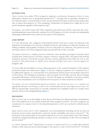
The brain MRI showed bilateral contrast-enhancing lesions in the cerebral hemispheres with moderate
perilesional edema and enhancement [Figure 1]. Because malignant lymphoma was considered first in
the differential diagnosis, no corticosteroids were administered and the patient was promptly referred for
an FNA biopsy. The histological examination, which included luxol fast blue staining, showed findings
compatible with lesions of demyelinating nature. The anti-TNF agent was considered as a potential causative
factor for the demyelination, thus was withdrawn from the patient. No other treatment was initiated.
The patient, who has eventually been placed on interferon, was followed up every 2 to 3 months with
appropriate laboratory monitoring, as well as with complete physical and neurological examinations. An
MRI was repeated after a mean period of 6 months. Symptoms were completely resolved in 2 months with a
dramatic retrieval of the imaging findings [Figure 2].
DISCUSSION
This is the first case, to our knowledge, to show that an FNA biopsy has differentiated brain demyelination
from CNS lymphoma in an RA patient, treated with TNFα antagonist and MTX. The TNFα antagonist was
considered as the causative factor for the demyelinating lesions, which were resolved by withdrawing the
medication.
Many adverse events, due to TNFα antagonists, have been published, including central nervous and
[4-6]
peripheral nervous system demyelination, transverse myelitis, retrobulbar optic neuritis and more . The
mechanism of this demyelination is not perfectly outlined, since it is not certain whether anti-TNFα blockers
unmask pre-existing demyelinating disorders or induce de novo demyelination of the CNS and peripheral
[1,7]
nervous system .
[5]
In a French national survey , demyelination occurred in a median period of 10.2 months after treatment
[8]
initiation, while etanercept was the reported agent in the majority of cases of CNS involvement . Of interest