
Page 128 - Read Online
P. 128
Page 4 of 5 Bogdos et al. Neuroimmunol Neuroinflammation 2018;5:19 I http://dx.doi.org/10.20517/2347-8659.2018.07
A B C D
E F G H
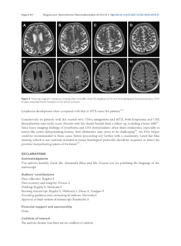
Figure 2. Follow up magnetic resonance imaging after 3 months. Axial T2-weighted (A-D) and fluid-attenuated inversion recovery (E-H)
images reveal significant remission of the lesions (arrows)
lymphoma development when compared with that in MTX-naive RA patients .
[12]
Conclusively, in patients with RA treated with TNFα antagonists and MTX, both lymphoma and CNS
[7]
demyelination may rarely occur. Patients with RA should benefit from a follow-up, including a brain MRI .
Since brain imaging findings of lymphoma and CNS demyelination often share similarities, especially in
[9]
tumor-like active-demyelinating lesions, their distinction may prove to be challenging . An FNA biopsy
could be recommended in these cases, before proceeding any further with a craniotomy. Luxol fast blue
staining (which is not routinely included in tumor histological protocols) should be requested to detect the
[13]
potential demyelinating nature of the lesion .
DECLARATIONS
Acknowledgments
The authors heartily thank Ms. Alexandra Bitza and Ms. Suyeon Lee for polishing the language of the
manuscript.
Authors’ contributions
Data collection: Bogdos E
Data accuracy and integrity: Drosos A
Drafting: Bogdos E, Markoula S
Revising manuscript: Bogdos E, Markoula S, Zikou A, Voulgari P
Providing guidance and connecting all authors: Markoula S
Approval of final version of manuscript: Konitsiotis S
Financial support and sponsorship
None.
Conflicts of interest
The authors declare that there are no conflicts of interest.