
Page 132 - Read Online
P. 132
Verma et al. Neuroimmunol Neuroinflammation 2018;5:20 I http://dx.doi.org/10.20517/2347-8659.2017.65 Page 3 of 6
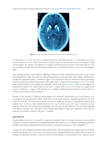
Figure 2. Computerized tomography scan of the head revealed bilateral basal ganglia
the duration of 6 months. She has 6-7 attacks/month and each attack last for 3-4 min without aura with
postictal state for 30 min. There was no past or family history of epilepsy. She was on antiepileptic drugs
carbamezapine 800 mg/day and clobazam 20 mg/day started by local practitioner. The patient did not have
any symptoms of hypocalcemia. Her general examination was normal and there were no focal neurological
signs.
Signs of hypocalcaemia were elicited by inflating a cuff above systolic blood pressure over the arm for 2 min,
provoking flexion of the wrist and metacarpophalangeal joints, hyperextension of the fingers, and flexion of
the thumb (carpopedal spasm - Trousseau’s sign), and by tapping the face just anterior to the ear, provoking
twitching of the ipsilateral facial muscles (Chovstek’s sign). Computerized tomography (CT) scan of the
head revealed bilateral basal ganglia and cerebellar calcification [Figures 2 and 3]. She was investigated for
hypoparathyroidism. Her serum calcium level was 5.9 mg/dL with a serum PTH level of 2.8 pg/mL and
serum phosphorus 7.2 mg/dL. EEG showed runs of higher amplitude theta and delta activity with very
marked response during hyperventilation.
Because of the presence of bilateral basal ganglia and cerebellar calcification on CT, the patients were
investigated for hypoparathyroidism. Both patients had hypocalcemia, hyperphosphatemia with low
serum PTH levels. The biochemical profile favored the diagnosis of primary hypoparathyroidism. Both
patients were started on daily supplementation of 0.5 µg of calcitriol and 1000 mg of elemental calcium
(calcium carbonate). The patients were discharged with oral calcium and vitamin D. The biochemical profile
normalized, seizures were controlled, and eventually antiepileptic drugs were withdrawn. They did not
experience any further seizures in the next 12 months.
DISCUSSION
Hypoparathyroidism can be caused by congenital disorders (like Di George syndrome, mitochondrial
cytopathies), receptor insensitivity (pseudohypoparathyroidism type Ia–c, II), surgery, autoimmune disorders
[2,3]
(familial autoimmune polyglandular syndrome type I), or hemochromatosis, or can be idiopathic .
Acquired chronic hypoparathyroidism is generally the after effect of unintentional surgical removal of all the
parathyroid glands. Even rarer causes of acquired chronic hypoparathyroidism include radiation-induced
damage and glandular damage in patients with hemochromatosis or hemosiderosis after repeated blood