
Page 127 - Read Online
P. 127
Bogdos et al. Neuroimmunol Neuroinflammation 2018;5:19 I http://dx.doi.org/10.20517/2347-8659.2018.07 Page 3 of 5
A B C
D E F
G H I
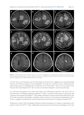
Figure 1. Axial fluid-attenuated inversion recovery (A-C) and T2-weighted (D-F) images show multiple large juxtacortical and
periventricular hyperintense lesions (arrows). Axial post-contrast T1-weighted images (G-I) demonstrate that the lesions abnormally
enhance, suggesting an active demyelinating process (curved arrows)
is the fact that, in most reported cases, the chronology of clinical events is suggestive of a causal relationship
between anti-TNFα and induction of demyelination, but only few cases have shown a definite positive
[5]
relationship, proven by rechallenge after re-admission of anti-TNF agents . There is only one report in the
[9]
literature of demyelinating lesions , that is proven to be demyelinating after craniectomy and biopsy.
It is well known that patients with certain autoimmune and inflammatory disorders, such as RA, have an
[10]
increased risk of developing malignant lymphoma with an overall two-fold increase in lymphoma risk
[3]
compared with the general population . The possible mechanisms for this increased risk include the fact that
RA results in persistent immunologic stimulation (which may lead to clonal selection and predispose CD5+
[3]
B cells to malignant transformation), and decreased number and function of T-suppressor lymphocytes .
Furthermore, mature T/NK-cell lymphoproliferative diseases development is a frequent complication in RA
[11]
patients treated with MTX , which additionally may shorten the interval between the diagnosis of RA and