
Page 19 - Read Online
P. 19
Turner et al. LPS preconditioning neuroprotective
GFAP expression was quantified to measure 0.05). Again, no difference was observed between
astrocyte activation and was significantly different LPS-treated and sham-injured rats at this time point
between experimental groups (F 3,32 = 6.30; P < 0.05) (q = 0.45; P > 0.05). Importantly, there was a strong
[Figure 6]. LPS preconditioning significantly reduced correlation between GFAP and OSMR expression
GFAP expression after TBI as shown by one-way evidenced by the yellow overlay (overlap coefficient
ANOVA and post-hoc Tukey’s test (q = 4.44; P < r = 0.722), indicating a high degree of overlap within
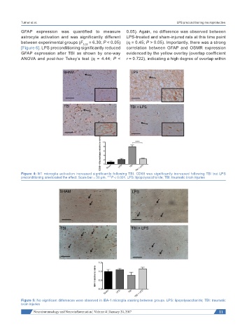
Figure 4: M1 microglia activation increased significantly following TBI. CD68 was significantly increased following TBI but LPS
preconditioning ameliorated the effect. Scale bar = 50 µm. ***P < 0.001. LPS: lipopolysaccharide; TBI: traumatic brain injuries
Figure 5: No significant differences were observed in IBA-1 microglia staining between groups. LPS: lipopolysaccharide; TBI: traumatic
brain injuries
Neuroimmunology and Neuroinflammation ¦ Volume 4 ¦ January 20, 2017 11