
Page 168 - Read Online
P. 168
Liu et al. Neuroinflammation in ischemic stroke
essential innate immune modulators and conduct an factors, which prevent inflammation and improve injury
array of biological processes. [43] [Figure 2]. M1 microglia tends to induce neuronal
[63]
cell death. Recent research has demonstrated that
The neuroinflammation process is decided by the the M1 phenotype microglia can be switched to the
scene, duration and course of the neurological M2 phenotype. One study has shown that HIV-
[64]
insult. Neuroinflammation can perform function associated dementia initiates and maintains M1
[44]
that are either supportive or destructive by which is phenotype microglia in the CD40 ligation by CD40L
determined by the immune signals relayed to the and TNFα. These microglia may later switch microglia
[65]
CNS. The nature of neuroinflammatory function to the M2 phenotype via up-regulation of CD45. In
can depend on the conditions and the intensity a pathological condition, the corresponding stimuli
and duration of inflammation. The positive role may active microglia and cause them to change
[45]
associated with neuroinflammation is only present their shape and function and initiate phagocytosis.
[66]
for a brief, controlled inflammatory situations and Microglia works in close association with astrocytes
responses and this can be considered as performing to release cytokines that lead to a cascade of events
a protective function to the host organism. [46-48] For which can modulate the neuroinflammatory respond.
example, during low transient inflammation that may Meanwhile, the microglia cells produce and release
occur during infections, the immune cell signals to the excitotoxic metabolites that can damage surrounding
brain by increasing the expression of interleukin (IL)- tissue. Sometimes a short-term neuroinflammatory
1 cytokine, this then increasing the ‘survellience’ role response is likely good for recovering the damages
[67]
of glia cells in the brain if infected. [49,50] The transient or infected tissue. On the contrary, a long period
inflammation of traumatic CNS injury, following the of time neuroinflammatory process may damage the
expression of IL-4, has been shown to promote injury surrounding brain tissue. [68]
recovery and axonal regrowth. [51,52] On the contrary,
the negative aspects of neuroinflammation mainly ROLE OF MICROGLIA IN
represent maladaptive inflammatory responses. [53,54] NEUROINFLAMMATION AFTER STROKE
The common characteristics of this aspect is
increasing, supraphysiological production of cytokines Neuroinflammation occurs in different types of
[IL-1 and tumor necrosis factor (TNF)], ROS, and other brain injuries including ischemic stroke. Ischemic
inflammatory mediators including inducible nitric oxide stroke mediated brain injury results in necrosis
synthase. These markers are highly evident in the and apoptosis. [69-71] The damaged cells and debris
[55]
high traumatic CNS, giving rise to collateral damage. induces neuroinflammation in areas in and around the
[56]
Following the acute phase of CNS trauma, the IL-1 ischemic injury in the brain. [72] Ischemia-induced cell
and IL-6 drive a low-level and chronic inflammatory debris and increased ROS lead to neuroinflammation
response, leading to cognitive impairments and by activating resident microglia and astrocytes
reduced neuronal plasticity. [57] as well as attracting infiltrating leukocytes from
circulating blood. [73] These cells increase major
MICROGLIA AND NEUROINFLAMMATION
Microglia are the innate immune cells of the CNS,
and are key modulators of the immune response in
the brain. Microglia is considered as the resident
[37]
macrophage in the brain and the initial responders
to tissue damage. Microglia express receptors that
[58]
respond to various stimuli that may as a consequence
result in there activation. A large number of studies
[59]
indicate that microglia expresses different proteins and
cytokines that display different role to express different
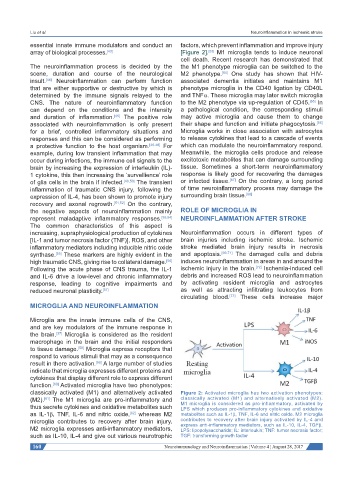
function. Activated microglia have two phenotypes:
[60]
classically activated (M1) and alternatively activated Figure 2: Activated microglia has two activation phenotypes:
(M2). The M1 microglia are pro-inflammatory and classically activated (M1) and alternatively activated (M2).
[61]
thus secrete cytokines and oxidative metabolites such M1 microglia is considered as pro-inflammatory, activated by
LPS which produces pro-inflammatory cytokines and oxidative
as IL-1β, TNF, IL-6 and nitric oxide, whereas M2 metabolites such as IL-1β, TNF, IL-6 and nitric oxide. M2 microglia
[62]
microglia contributes to recovery after brain injury. contributes to recovery after brain injury activated by IL-4 and
express anti-inflammatory mediators, such as IL-10, IL-4, TGFβ.
M2 microglia expresses anti-inflammatory mediators, LPS: lipopolysaccharide; IL: interleukin; TNF: tumor necrosis factor;
such as IL-10, IL-4 and give out various neurotrophic TGF: transforming growth factor
160 Neuroimmunology and Neuroinflammation ¦ Volume 4 ¦ August 28, 2017