
Page 164 - Read Online
P. 164
Wang et al. Cytology of cerebrospinal and superficial siderosis
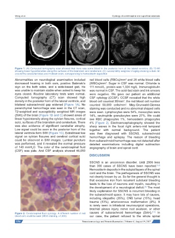
Figure 1: (A) Computed tomography scan showed that there was some blood in the posterior horn of the lateral ventricle; (B) T2-WI
showing linear hypointensities along the surfaces of the brainstem and cerebellum; (C) susceptibility weighted imaging showing a dark rim
around the cerebellar lobes and midbrain brain, corresponding to hemosiderin deposition
Abnormalities on neurological examination included red blood cells (RBCs)/mm and 28 white blood cells
3
decreased hearing in both ears, positive Babinski’s (WBCs)/mm . Sugar in CSF was normal. Chloride is
3
sign on the both sides, and a wide-based gait. He 111 mmol/L, protein was 1,324 mg/L. Immunoglobulin
was unable to maintain stable when asked to keep his was normal in CSF. The acid-fast stain and ink smears
eyes closed. Routine laboratory tests were normal. were negative. We gave our patient an additional
Computed tomography (CT) scan showed high CSF cytology (CCSF). CCSF revealed that the white
density in the posterior horn of the lateral ventricle, and blood cell counted 30/mm , the red blood cell number
3
bilateral subarachnoid gap widened [Figure 1A]. No counted 50,000 cells/mm . May-Grunwald-Giemsa
3
parenchymal hemorrhage was seen in the CT scan, staining was conducted and no abnormal shaped cells
T2-weighted and susceptibility weighted MR images were seen. Lymphocytes were 54%, monocytes were
(SWI) of the brain [Figure 1B and C] showed areas of 14%, neutrophile granulocytes were 27%. We could
linear hypointensity along the sylvian fissures, cortical see RBC phagocytes 1%, hemosiderin phagocytes
sulci, surfaces of the brainstem and cerebellum. There 4% [Figure 2]. Electroencephalography showed the
was also evidence of significant cerebellar atrophy. sharp waves in the focal right antero-mid temporal
Low signal could be seen in the posterior horn of the together with normal background. The patient
lateral ventricle form SWI [Figure 1C]. Extrallinear low was then diagnosed with SSCNS, subarachnoid
signal on sylvian fissures and cerebral cortical sulci hemorrhage and epilepsy. The reason of bleeding
could be observed in SWI images. Lumbar puncture from subarachnoid hemorrhage was not detected after
was performed, and it revealed the normal pressure detailed examinations including digital subtraction
of 140 mmH O. The color of the cerebrospinal fluid angiography of brain and spinal cord.
2
(CSF) was pale. And CSF analysis showed 46,050
DISCUSSION
SSCNS is an uncommon disorder. Until 2006 less
than 300 cases of SSCNS have been reported. [1,2]
Hemosiderin deposits in the subpial layers of the spinal
cord and the brain. The pathogenesis of SSCNS was
not cleanly known by us. So far the general thought is
that excessive iron from recurrent subdural bleeding
leads to the loss of neurons and myelin, resulting in
the development of a neurological deficit. The most
[3]
likely explanation for SSCNS is recurrent bleeding in
the subarachnoid space. It may have lots of causes,
including idiopathic (35%), CNS tumor (15%), head
trauma (13%), arteriovenous malformation (9%). It
is rarely seen in intradural neurosurgical operations,
brachial plexus injury, nerve root avulsion, or other
Figure 2: Cerebrospinal fluid cytology. A different number of red causes of subarachnoid hemorrhage (SAH). [4-6] In
blood cells could be seen (MGG staining, ×1,000) our case, the patient refused to the whole spinal
156 Neuroimmunology and Neuroinflammation ¦ Volume 4 ¦ August 09, 2017