
Page 76 - Read Online
P. 76
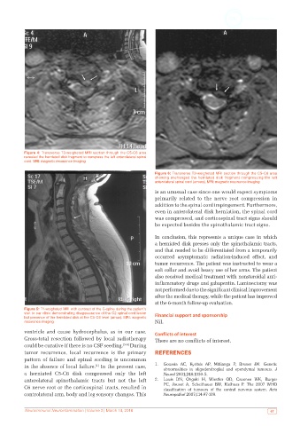
Figure 4: Transverse T2-weigheted MRI section through the C5-C6 area
           revealed the herniated disk fragment to compress the left anterolateral spinal
           cord. MRI: magnetic resonance imaging

                                                               Figure 6: Transverse T2-weigheted MRI section through the C5-C6 area
                                                               showing unchanged the herniated disk fragment compressing the left
                                                               anterolateral spinal cord (arrows). MRI: magnetic resonance imaging

                                                               is an unusual case since one would expect symptoms
                                                               primarily related to the nerve root compression in
                                                               addition to the spinal cord impingement. Furthermore,
                                                               even in anterolateral disk herniation, the spinal cord
                                                               was compressed, and corticospinal tract signs should
                                                               be expected besides the spinothalamic tract signs.

                                                               In conclusion, this represents a unique case in which
                                                               a herniated disk presses only the spinothalamic tracts,
                                                               and that needed to be differentiated from a temporarily
                                                               occurred asymptomatic radiation-induced effect, and
                                                               tumor recurrence. The patient was instructed to wear a
                                                               soft collar and avoid heavy use of her arms. The patient
                                                               also received medical treatment with nonsteroidal anti-
                                                               inflammatory drugs and gabapentin. Laminectomy was
                                                               not performed due to the significant clinical improvement
                                                               after the medical therapy, while the patient has improved
                                                               at the 6-month follow-up evaluation.
           Figure 5: T1-weigheted MRI with contrast of the C-spine during the patient’s
           visit to our clinic demonstrating disappearance of the C2 spinal cord lesion   Financial support and sponsorship
           but presence of the herniated disk at the C5-C6 level (arrow). MRI: magnetic
           resonance imaging                                   Nil.
           ventricle and cause hydrocephalus, as in our case.   Conflicts of interest
           Gross-total resection followed by local radiotherapy   There are no conflicts of interest.
           could be curative if there is no CSF seeding. [3-5]  During
           tumor recurrence, local recurrence is the primary   REFERENCES
           pattern of failure and spinal seeding is uncommon
           in the absence of local failure.  In the present case,   1.   Goussia AC, Kyritsis AP, Mitlianga P, Bruner JM. Genetic
                                       [1]
                                                                  abnormalities in oligodendroglial and ependymal tumours.  J
           a herniated C5-C6 disk compressed only the left        Neurol 2001;248:1030-5.
           anterolateral  spinothalamic  tracts  but  not the left   2.   Louis  DN,  Ohgaki H,  Wiestler OD,  Cavenee WK,  Burger
           C6 nerve root or the corticospinal tracts, resulted in   PC, Jouvet A, Scheithauer BW, Kleihues P. The 2007 WHO
                                                                  classification of  tumours  of  the central nervous  system.  Acta
           controlateral arm, body and leg sensory changes. This   Neuropathol 2007;114:97-109.


           Neuroimmunol Neuroinfammation | Volume 3 | March 14, 2016                                        67