
Page 75 - Read Online
P. 75
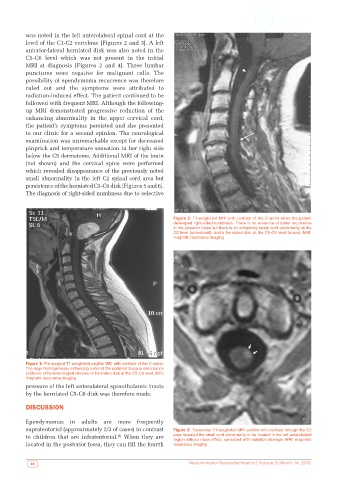
was noted in the left anterolateral spinal cord at the
          level of the C1-C2 vertebrae [Figures 2 and 3]. A left
          anterior-lateral herniated disk was also noted in the
          C5-C6 level which was not present in the initial
          MRI at diagnosis [Figures 2 and 4]. Three lumbar
          punctures were negative for malignant cells. The
          possibility of ependymoma recurrence was therefore
          ruled out and the symptoms were attributed to
          radiation-induced effect. The patient continued to be
          followed with frequent MRI. Although the following-
          up MRI demonstrated progressive reduction of the
          enhancing abnormality in the upper cervical cord,
          the patient’s  symptoms  persisted  and  she presented
          to our clinic for a second opinion. The neurological
          examination was unremarkable except for decreased
          pinprick and temperature sensation in her right side
          below the C5 dermatome. Additional MRI of the brain
          (not  shown)  and  the  cervical  spine  were  performed
          which revealed disappearance of the previously noted
          small abnormality in the left C2 spinal cord area but
          persistence of the herniated C5-C6 disk [Figures 5 and 6].
          The diagnosis of right-sided numbness due to selective


                                                              Figure 2: T1-weigheted MRI with contrast of the C-spine when the patient
                                                              developed right-sided numbness. There is no evidence of tumor recurrence
                                                              in the posterior fossa but there is an enhancing spinal cord abnormality at the
                                                              C2 level (arrowhead), and a herniated disk at the C5-C6 level (arrow). MRI:
                                                              magnetic resonance imaging


          Figure 1: Pre-surgical T1-weigheted sagittal MRI with contrast of the C-spine.
          The large homogenously enhancing tumor of the posterior fossa is seen but no
          evidence of leptomeningeal disease or herniated disk at the C5-C6 level. MRI:
          magnetic resonance imaging
          pressure of the left anterolateral spinothalamic tracts
          by the herniated C5-C6 disk was therefore made.
          DISCUSSION


          Ependymomas in adults are more frequently
          supratentorial (approximately 2/3 of cases) in contrast   Figure 3: Transverse T1-weigheted MRI section with contrast through the C2
          to children  that are infratentorial.   When  they  are   area revealed the small cord abnormality to be located in the left anterolateral
                                          [1]
                                                              region without mass effect, consisted with radiation damage. MRI: magnetic
          located in the posterior fossa, they can fill the fourth   resonance imaging

            66                                                     Neuroimmunol Neuroinfammation | Volume 3 | March 14, 2016