
Page 171 - Read Online
P. 171
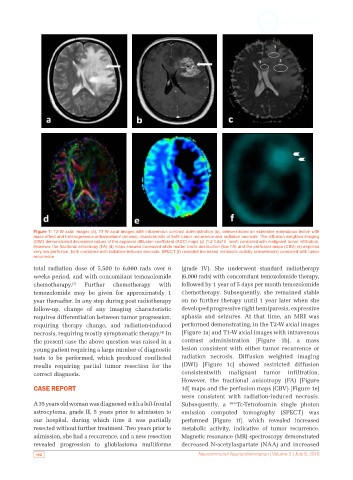
Figure 1: T2-W axial images (a), T1-W axial images with intravenous contrast administration (b), demonstrated an extensive edematous lesion with
            mass effect and heterogeneous enhancement (arrows), characteristic of both tumor recurrence and radiation necrosis. The diffusion weighted imaging
                                                                              ³
            (DWI) demonstrated decreased values of the apparent diffusion coefficient (ADC) maps (c) (1.2-1.8x10¯ mm²) consisted with malignant tumor infiltration.
            However, the fractional anisotropy (FA) (d) maps showed increased white matter tracts destruction (low FA) and the perfusion maps (CBV) (e) depicted
            very low perfusion, both consisted with radiation-induced necrosis. SPECT (f) revealed increased metabolic activity (arrowheads) consisted with tumor
            recurrence
            total radiation dose of 5,500 to 6,000 rads over 6   (grade IV). She underwent standard radiotherapy
            weeks period, and with concomitant temozolomide   (6,000 rads) with concomitant temozolomide therapy,
            chemotherapy. [3]  Further  chemotherapy   with   followed by 1 year of 5 days per month temozolomide
            temozolomide may be given for approximately 1     chemotherapy. Subsequently, she remained stable
            year thereafter. In any step during post radiotherapy   on no further therapy until 1 year later when she
            follow-up, change of any imaging characteristic   developed progressive right hemiparesis, expressive
            requires differentiation between tumor progression,   aphasia and seizures. At that time, an MRI was
            requiring  therapy  change,  and  radiation-induced   performed demonstrating, in the T2-W axial images
            necrosis, requiring mostly symptomatic therapy.  In   [Figure 1a] and T1-W axial images with intravenous
                                                       [4]
            the present case the above question was raised in a   contrast administration [Figure 1b], a mass
            young patient requiring a large number of diagnostic   lesion consistent with either tumor recurrence or
            tests to be performed, which produced conflicted   radiation necrosis. Diffusion weighted imaging
            results requiring  partial tumor resection for the   (DWI)  [Figure  1c]  showed  restricted  diffusion
            correct diagnosis.                                consistentwith malignant tumor infiltration.
                                                              However, the fractional anisotropy (FA) [Figure
            CASE REPORT                                       1d] maps and the perfusion maps (CBV) [Figure 1e]
                                                              were consistent with radiation-induced necrosis.
            A 35 years old woman was diagnosed with a left-frontal   Subsequently, a   99m Tc-Tetrofosmin single photon
            astrocytoma, grade II, 5 years prior to admission to   emission computed tomography (SPECT) was
            our hospital, during which time it was partially   performed [Figure 1f], which revealed increased
            resected without further treatment. Two years prior to   metabolic activity, indicative of tumor recurrence.
            admission, she had a recurrence, and a new resection   Magnetic resonance (MR) spectroscopy demonstrated
            revealed  progression  to  glioblastoma  multiforme   decreased N-acetylaspartate (NAA) and increased
             162                                                    Neuroimmunol Neuroinflammation | Volume 3 | July 8, 2016