
Page 166 - Read Online
P. 166
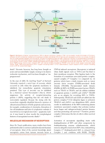
Figure 1: Mechanism of tumour necrosis factor-α (TNF-α) induced necroptosis. Binding of TNF-α to its receptor results in formation of Complex I.
            Activation of cIAP and tartrate resistant acid phosphatase activates downstream NF-κB signaling and subsequently promote cell survival. Complex II
            acts as a switch between apoptosis and necroptosis. Activation of caspase-8 guides the cells to apoptosis. Inhibition of caspase-8 leads to formation of
            a necrosome. Membrane translocation of phosphrylated MLKL disrupts cell membrane. The mechanisms underlying the lysis of cytoplasmic contents
            during necrosis are still unclear. DAMPs: damage associated molecular patterns; MLKL: mixed lineage kinase domain-like protein; TNF: tumour necrosis
            factor; TNFR: tumour necrosis factor receptor; cIAP: calf intestinal alkaline phosphatise; TRADD: tumor necrosis factor receptor associated death
            domain; TRAF: TNFR-associated factors; RIP1: receptor-interacting protein 1; CYLD: cylindromatosis; Casp8: caspase-8; RIP3: receptor-interacting
            protein 3; NF-kB: nuclear factor kappa.
            death”. Necrosis, however, has long been thought as   (TNF-α) induced necroptosis. Necroptosis is initiated
            acute and uncontrollable, largely owning to its elusive   when death signals such as TNF-α and Fas bind to
            molecular mechanism, and thus been thought as “un-  their membrane receptors. This ligation leads to the
            programmed”.                                      formation of a membrane associated protein complex,
                                                              named complex I.  Complex I is composed by: (1)
                                                                              [4]
            In the year of 2005, Dr. Jun-Ying Yuan  at Harvard   proteins which have a death domain such as tumor
                                               [2]
            University  reported  a  novel  type  of  necrosis,  which   necrosis factor receptor (TNFR)-associated death
            occurred in cells when the apoptosis machinery is   domain (TRADD), Fas-associated death domain
            inhibited, but extracellular apoptotic stimulation   (FADD); (2) RIP1; (3) TNFR-associated factors (TRAF),
            persisted. This type of necrosis can be inhibited   such as TRAF2 or TRAF5, and (4) cellular inhibitor
            by a chemical named Necrostatin-1 (Nec-1), which   of apoptosis protein 1 (cIAP1) and cIAP2.  TRADD
                                                                                                    [5]
            suppresses the activity of receptor-interacting   acts as an adaptor for recruiting RIP1 to TNFR1.
            protein 1 (RIP1), suggesting that the cell death is   Subsequently, TRAF2/3/5 and cIAPs are added into the
            molecularly regulated. Because the dying cells the   protein complex.  If E3 ubiquitin ligase is activated,
                                                                             [6]
            researchers originally identified showed a mixture of   TRAF2/5 and cIAP1/2 can ubiquitinate RIP1, which
            ultrastructural features of both apoptosis and necrosis,   results in stabilization of the RIP1-containing plasma
            for example, condensation of chromatin, disruption of   membrane associated complex that activates nuclear
            the cell membrane and lysis of cytoplasmic contents, it   factor kappa and mitogen-activated protein kinases,
            was termed as necroptosis (necrosis + apoptosis). Later   and thus promoting cell survival.  Therefore, protein
                                                                                           [7]
            on, it was found to show mostly the morphological   complex I determines the fate of cells to either survival
            features of unregulated necrotic death. [3]       or death. [8]
            MOLECULAR MECHANISM OF NECROPTOSIS                Activation  of  necroptosis  signalling  starts  with
                                                              deubiquitination of RIP1 and other components by
            Since Dr. Yuan’s publication, many studies have been   deubiquitinating enzyme  cylindromatosis, which
            performed on the occurrence and molecular mechanism   removes ubiquitin chains from RIP1, thus, destabilizing
            of necroptosis. Most of the current knowledge about   Complex I.  Deubiquitinated RIP1 is released from
                                                                        [9]
            necroptosis comes from tumour necrosis factor  α   Complex I and combines with FADD, TRADD,
            Neuroimmunol Neuroinflammation | Volume 3 | July 8, 2016                                      157