
Page 172 - Read Online
P. 172
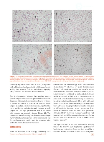
Figure 2: (a) Histological examination revealed a neoplasm with highly pleomorphic cells exhibiting several mitoses (arrows); (b) microvascular
            proliferation was also evident, a common finding in high grade gliomas (arrows); (c) some areas of the biopsied tumor showed evidence of radiation-
            induced changes, such as necrosis (arrows) and vascular changes such as telangiectasia and hyalinization of vessel walls adjacent to areas of
            hypercellular tumor tissue (arrows); (d) the proliferation marker MIB1 (Ki67) was expressed in a high proportion of tumor cells (arrows).
            choline (Cho) with ratio Cho/NAA > 3.63, compatible   combination of radiotherapy with temozolomide
            with infiltration of malignant cells with high metabolic   chemotherapy  followed by plain temozolomide
                                                                           [3]
            activity (not shown). Positron emission tomography   therapy, glioblastoma multiforme usually  recurs
                                                                                            [5]
            (PET) scan was not available atour institution.   within the first year from therapy.  However, at that
                                                              point  it  may  be  difficult  to  differentiate  between
            Due to discrepancy between the imaging tests, a   radiation necrosis of the tumor vs. tumor recurrence,
            partial surgical resection was performed for accurate   since both conditions behave similarly on traditional
            diagnosis. Histological examination showed evidence   imaging modalities (Standard CT or MRI with and
            of tumor recurrence in most of the resected tissue   without IV contrast administration).  In these cases,
                                                                                              [6]
            [Figure 2a and 2b]. There were some small parts of the   more advanced imaging modalities can be employed
            tumor  exhibiting  radiation-induced  changes  as  well   to differentiate between tumor recurrence from
            [Figure 2c]. The high proliferation rate of the tumor   radiation necrosis,  such as  PET  and perfusion/
                                                                                             [7]
            cells denoted an aggressive tumor [Figure 2d]. The   diffusion MRI.  However, in many countries, PET
                                                                            [8]
            patient was started on daily low dose temozolomide (50   is not widely available, necessitating the use of other
            mg/m  of body surface per os) administration and oral   nuclear medicine modalities such as SPECT with
                 2
            dexamethasone at 6 mg/day, and she remained alive   various tracers. [9]
            and stable 6 months after the operation.
                                                              MR  spectroscopy  is  another  alternative  imaging
            DISCUSSION                                        method to improve our diagnostic capabilities in
                                                              brain  tumor  evaluation,  however,  this  modality  is
            After  the  standard  initial  therapy,  consisting  of  a   still not widely available.  NAA is an acetylated
                                                                                      [10]
            Neuroimmunol Neuroinflammation | Volume 3 | July 8, 2016                                      163