
Page 87 - Read Online
P. 87
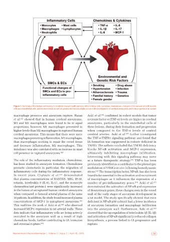
Figure 1: Summary of the factors contributing to cerebral aneurysm pathogenesis. Inflammatory cells, cytokines, chemokines, changes to the vascular smooth muscle
           cells and endothelial cells, and environmental as well as genetic risk factors all play a role in the development of cerebral aneurysms and their progression to rupture

           macrophage presence and aneurysm rupture. Hasan    Aoki et al. [25]  confirmed in rodent models that tumor
           et al. [22]  showed that in human cerebral aneurysms,   necrosis factor-α (TNF-α) levels are higher in cerebral
           M1 and M2 macrophages were found to be in equal    aneurysms, particularly in the endothelial cells of
           proportions; however, M1 macrophages presented in   these lesions, during their formation and progression
           higher levels than M2 macrophages in ruptured human   when compared to the TNF-α levels of control
           cerebral aneurysms. This means that there were more   cerebral arteries. Aoki et  al. [25]  further investigated

           macrophages promoting inflammation, M1 macrophages,   the TNF-α-TNFR1 signaling pathway and found that
           than macrophages working to repair the vessel tissue   IA formation was suppressed in rodents deficient in
           and decrease inflammation, M2 macrophages. This    TNFR1. The authors concluded that TNFR1 deficiency
           imbalance was also correlated with an increase in mast   blocks NF-κB activation and MCP-1 expression,
           cell presence in ruptured aneurysms. [22]          ultimately inhibiting macrophage infiltration.
                                                              Intervening with this signaling pathway may serve
           The role of the inflammatory mediators, chemokines,   as a future therapeutic strategy. [25]  TNF-α has been
           has been studied in aneurysm formation. Chemokines   previously identified as a contributor to the phenotypic
           promote chemotaxis in particular, the migration of   modulation of VSMCs in vivo following hemodynamic
           inflammatory cells during the inflammatory response.   stress. [26]  The transcription factor, NF-κB, has also been
           In recent years, Chalouhi  et  al. [23]  demonstrated   found to be essential in the activation and recruitment
           that plasma concentrations of RANTES, MIG, IP-10,   of macrophages as it influences the expression of a
           eotaxin, interleukin 8 (IL-8), IL-17, and the monocyte   number of pro-inflammatory genes. [5,6]  Aoki et al. [27]
           chemoattractant protein-1 were significantly increased   demonstrated the activation of NF-κB and expression
           in the lumen of unruptured human cerebral aneurysms   of downstream genes; these changes seen in the vessel
           when compared to femoral arterial plasma of the same   wall of the early stages of aneurysm development in
           16 patients. In addition, the study found increased plasma   a rat model. The study specifically showed that mice
                                                         [23]
           concentrations of MCP-1 in unruptured aneurysms.    deficient in NF-κB p50 subunit had a lower incidence
                                            [24]
           This reaffirms the work of Aoki et al.  who observed   of aneurysm formation and macrophage infiltration
           increased MCP-1 expression in rat arterial walls. These   into the aneurysm wall. Furthermore, Aoki et al. [28]
           data indicate that inflammatory cells are being actively   showed that the up-regulation of interleukin-1β (IL-1β)
           recruited to the aneurysm wall as a result of high   and activation of NF-κB significantly reduced collagen
           chemokine levels, further contributing to IA formation   biosynthesis, a process linked to IA progression and
           and eventual rupture. [23]                         rupture.


          Neuroimmunol Neuroinflammation | Volume 2 | Issue 2 | April 15, 2015                              79