
Page 65 - Read Online
P. 65
+HPRG\QDPLF VKHDU VWUHVV
7KH ELIXUFDWLRQ RI FHUHEUDO DUWHU\
$FWLYDWLRQ RI 71) Į
3KRVSKRU\ODWLRQ E\ ,.. DQG VHFHVVLRQ
$FWLYDWLRQ RI 1) N%
1XFOHDU WUDQVORFDWLRQ
$FWLYDWLRQ RI LQIODPPDWRU\ UHODWHG UHDFWLRQ
([SUHVVLRQ RI 0&3
,QILOWUDWLRQ RI PDFURSKDJH
$FWLYDWLRQ RI L126 $FWLYDWLRQ RI
,/ ȕ 003V
$SRSWRVLV RI &OHDYDJH RI
60&V (&0
)UDJPHQWDWLRQ RI LQWHUQDO HODVWLF ODPLQDH
7KLQQLQJ RI PHGLD
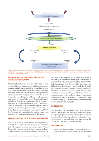
Figure 1: Factors associated with the formation of cerebral aneurysms. TNF-α: tumor necrosis factor-alpha; IKK: inhibitor of kappa kinase; NF-κB: nuclear factor-
kappa B; MCP-1: monocyte chemoattractant protein-1; iNOS: inducible nitric oxide synthase; IL-1β: interleukin-1β; MMPs: matrix metalloproteinases; SMCs: smooth
muscle cells; ECM: extracellular matrix
MECHANISMS OF CEREBRAL ANEURYSM wall around the ruptured site is immensely thin and
FORMATION IN HUMANS covered by a thrombosed fibrin plug. Infiltration of
inflammatory cells such as neutrophils, lymphocytes,
Aneurysm formation has been shown to occur at sites and macrophages into the adventitia also occurs. In
of constant hemodynamic stress both in humans and in addition, complement and immunoglobulin deposits
experimentally induced models of cerebral aneurysm. form between the medial vascular smooth muscle and
This means that hemodynamic stress initiates early stage adventitia. [13] Loss of vascular smooth muscle cells,
aneurysm formation. Aneurysm formation progresses thinning of collagen fibers, and inflammation are
when degenerative changes exceed vessel repair due more prominent in the walls of ruptured aneurysms
to vascular remodeling. In addition to inflammation compared to unruptured ones. [30] Therefore, these
and apoptosis, extracellular matrix decomposition and factors seem to play an important role in the weakening
endothelial dysfunction play critical roles in aneurysm of aneurysmal walls [Figure 1].
formation. It has been found that gene expression in
cerebral aneurysms may be linked to specific genetic CONCLUSION
regions. Furthermore, genetic regions that promote
cerebral aneurysm formation are also linked to AAAs. [29] Inflammatory cells have been found in the walls of
The factor analysis which affected to cerebral aneurysmal cerebral aneurysms, and several inflammatory factors
formation in human would progress in the future. are reported to play crucial roles in cerebral aneurysm
formation. Possible therapeutic interventions to reduce
HISTOPATHOLOGY OF RUPTURED ANEURYSMS the formation of cerebral aneurysms may include the
inhibition of mediators of inflammation.
Loss of the internal elastic lamina and degeneration
of medial vascular smooth muscle are common REFERENCES
histopathological features of cerebral aneurysms. These
degenerative changes occur in the aneurysm wall and 1. Wiebers DO, Whisnant JP, Huston J 3rd, Meissner I, Brown RD Jr,
can lead to aneurysmal rupture. Following rupture, the Piepgras DG, Forbes G, Thielen K, Nichols D, O’Fallon W, Peacock J,
Neuroimmunol Neuroinflammation | Volume 2 | Issue 2 | April 15, 2015 57