
Page 60 - Read Online
P. 60
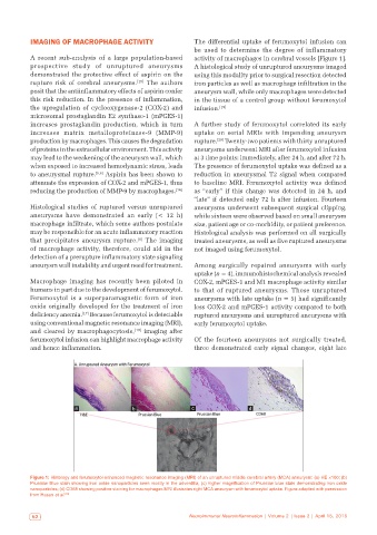
IMAGING OF MACROPHAGE ACTIVITY                     The differential uptake of ferumoxytol infusion can
                                                              be  used  to  determine  the  degree  of  inflammatory
           A recent sub-analysis of a large population-based   activity of macrophages in cerebral vessels [Figure 1].
           prospective study of unruptured aneurysms          A histological study of unruptured aneurysms imaged
           demonstrated the protective effect of aspirin on the   using this modality prior to surgical resection detected
           rupture risk of cerebral aneurysms. [15]  The authors   iron particles as well as macrophage infiltration in the
           posit that the antiinflammatory effects of aspirin confer   aneurysm wall, while only macrophages were detected
           this risk reduction. In the presence of inflammation,   in the tissue of a control group without ferumoxytol
           the upregulation of cyclooxygenase-2  (COX-2) and   infusion. [19]
           microsomal prostaglandin E2 synthase-1 (mPGES-1)
           increases prostaglandin production, which in turn   A further study of ferumoxytol correlated its early
           increases  matrix metalloproteinase-9  (MMP-9)     uptake on serial MRIs with impending aneurysm
           production by macrophages. This causes the degradation   rupture. [20]  Twenty-two patients with thirty unruptured
           of proteins in the extracellular environment. This activity   aneurysms underwent MRI after ferumoxytol infusion
           may lead to the weakening of the aneurysm wall, which   at 3 time points: immediately, after 24 h, and after 72 h.
           when exposed to increased hemodynamic stress, leads   The presence of ferumoxytol uptake was defined as a
           to aneurysmal rupture. [5,6]  Aspirin has been shown to   reduction in aneurysmal T2 signal when compared
           attenuate the expression of COX-2 and mPGES-1, thus   to  baseline MRI.  Ferumoxytol  activity was  defined
           reducing the production of MMP-9 by macrophages. [16]  as “early” if this change was detected in 24 h, and
                                                              “late” if detected only 72 h after infusion. Fourteen
           Histological studies of ruptured versus unruptured   aneurysms underwent subsequent surgical clipping,
           aneurysms have demonstrated an early  (< 12  h)    while sixteen were observed based on small aneurysm
           macrophage infiltrate, which some authors postulate   size, patient age or co-morbidity, or patient preference.
           may be responsible for an acute inflammatory reaction   Histological analysis was performed on all surgically
                                             [8]
           that precipitates aneurysm rupture.  The imaging   treated aneurysms, as well as five ruptured aneurysms
           of  macrophage  activity,  therefore,  could  aid  in  the   not imaged using ferumoxytol.
           detection of a prerupture inflammatory state signaling
           aneurysm wall instability and urgent need for treatment.  Among surgically repaired aneurysms with early
                                                              uptake (n = 4), immunohistochemical analysis revealed
           Macrophage imaging has recently been piloted in    COX-2, mPGES-1 and M1 macrophage activity similar
           humans in part due to the development of ferumoxytol.   to  that  of  ruptured  aneurysms.  Those  unruptured
           Ferumoxytol is a superparamagnetic form of iron    aneurysms with late uptake (n = 5) had significantly
           oxide originally developed for the treatment of iron   less COX-2 and mPGES-1 activity compared to both
                           [17]
           deficiency anemia.  Because ferumoxytol is detectable   ruptured aneurysms and unruptured aneurysms with
           using conventional magnetic resonance imaging (MRI),   early ferumoxytol uptake.
           and cleared by macrophagocytosis, [18]  imaging after
           ferumoxytol infusion can highlight macrophage activity   Of the fourteen aneurysms not surgically treated,
           and hence inflammation.                            three demonstrated early signal changes, eight late


                         a                 b                  c                 d


           Figure 1: Histology and ferumoxytol-enhanced magnetic resonance imaging (MRI) of an unruptured middle cerebral artery (MCA) aneurysm: (a) HE ×100; (b)
           Prussian Blue stain showing iron oxide nanoparticles seen mostly in the adventitia; (c) higher magnification of Prussian blue stain demonstrating iron oxide
           nanoparticles; (d) CD68 showing positive staining for macrophages MRI illustrates right MCA aneurysm with ferumoxytol uptake. Figure adapted with permission
           from Hasan et al. [19]


            52                                               Neuroimmunol Neuroinflammation | Volume 2 | Issue 2 | April 15, 2015