
Page 36 - Read Online
P. 36
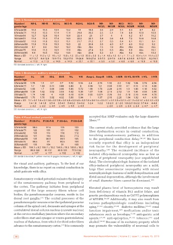
Table 2: Somatosensory evoked potentials
           Number/    N9‑L    N9‑R   N13‑L   N13‑R   N20‑L   N20‑R   N9‑    N9‑    N13‑   N13‑   N9‑     N9‑
           gender/age                                               N13/L  N13/R  N20/L N20/R   N20/L   N20/R
           3/female/68  10.3  10.3    13.9    13.2     19     18.5    3.6    2.9    5.1   5.3    8.7      8.2
           6/female/71  11.9  12.3    17.4    17.4    24.8    26.2    5.5    5.1    7.4   8.8    12.9    13.9
           10/male/64  12.7   12.8    16.4    16.8    22.4     23     3.7    4      6     6.2    9.7     10.2
           16/male/51  11.6   11.3    17.4    14.9    22.2    22.1    5.8    3.6    4.8   7.2    10.6    10.8
           23/male/62  11.6   11.8    15.3    15.1    21.7    21.2    3.7    3.3    6.4   6.1    10.1     9.4
           24/female/56  11.8  11.6   15.3    16.5     22     20.8    3.5    4.9    6.7   4.3    10.2     9.2
           28/female/58  8.7   8.6    16.2    16.2    Abs     Abs     7.5    7.6   Abs    Abs    Abs     Abs
           29/male/75  10.6   11.3    16.9    17.6    Abs     27.4    6.3    6.3   Abs    9.8    Abs     16.1
           30/female/65  9.9  10.5    15.2    14.6    Abs     23.8    5.3    4.1   Abs    9.2    Abs     13.3
           Mean ± SD  11 ± 1.2 11.2 ± 1.2 16 ± 1.2 15.8 ± 1.5 22 ± 1.9 22.9 ± 2.9 5 ± 1.4 4.6 ± 1.5 6.1 ± 1 7.1 ± 2 10.4 ± 1.4 11.4 ± 2.7
           Range     8.7‑12.7  8.6‑12.8  13.9‑17.4 13.2‑17.6  19‑24.8  18.5‑27.4 3.5‑7.5  2.9‑7.6  4.8‑7.4 4.3‑9.8  8.7‑12.9  8.2‑16.1
           Normal     ≤ 11.3  ≤ 11.3  ≤ 14.9  ≤ 14.9  ≤ 22.1  ≤ 22.1  ≤ 5.0  ≤ 5.0  ≤ 6.8  ≤ 6.8  ≤ 10.9  ≤ 10.9
           SD: standard deviation; L: left; R: right


           Table 3: Brainstem auditory evoked potentials
           Number/    I‑L    I‑R   III‑L  III‑R  V‑L   V‑R  Amp‑L Amp‑R I‑III/L  I‑III/R III‑V/L III‑V/R I‑V/L  I‑V/R
           gender/age
           3/female/68  1.78  1.5   3.7   3.7   5.52   5.54   2.4   2.16   1.92   2.2   1.82  1.84  3.74  4.04
           6/female/71  1.7  1.64  3.74   3.86  5.84    6     2.06   1.2   2.04   2.22  2.1  2.14  4.14   4.36
           23/male/62  1.68  1.7   3.96   3.86  5.86   5.72   1.98  1.76   2.28   2.16  1.9  1.86  4.18   4.02
           28/female/58  1.38  1.42  3.52  3.54  5.42  5.38   1.07  1.08   2.14   2.12  1.9  1.84  4.04   3.96
           29/male/75  1.74  1.48   4     4.08  6.18   6.24   2.05  0.974  2.26   2.6   2.18  2.16  4.44  4.76
           30/female/65  1.76  1.76  3.92  4.02  5.86  5.94    1    1.97   2.16   2.26  1.94  1.92  4.1   4.18
           Mean ± SD  1.7 ± 0.1 1.6 ± 0.1 3.8 ± 0.2 3.8 ± 0.2 5.8 ± 0.3 5.8 ± 0.3 1.8 ± 0.6 1.5 ± 0.5 2.1 ± 0.1 2.3 ± 0.2 2 ± 0.1 2 ± 0.2 4.1 ± 0.2 4.2 ± 0.3
           Range     1.4‑1.8  1.4‑1.8  3.5‑4  3.5‑4.1  5.4‑6.2  5.4‑6.2  1‑2.4  1‑2.2  1.9‑2.3  2.1‑2.6 1.8‑2.2 1.8‑2.2 3.7‑4.4  4‑4.8
           Normal    ≤ 2.2  ≤ 2.2  ≤ 4.5  ≤ 4.5  ≤ 6.5  ≤ 6.5             ≤ 2.6  ≤ 2.6  ≤ 2.4 ≤ 2.4  ≤ 4.7  ≤ 4.7
           SD: standard deviation; L: left; R: right


           Table 4: Visual evoked potentials                  accepted that SSEP evaluates only the large diameter
           Number/   P100‑P‑L  P100‑P‑R  P100‑G‑L  P100‑G‑R   fibers. [17]
           gender/age
           3/female/68  98.5     97.5      125      130       The current study provided evidence that the large
           6/female/71  122      131       131      132       fiber  dysfunction occurs  in  central conduction,
           23/male/62   103      114       116      112
           24/female/56  101     103      80.5      77.5      involving somatosensory pathway, in addition
           28/female/58  105     99.5     99.5      105       to the peripheral conduction delay. [16,18]  We have
           29/male/75   99.5     91       90.5      104       recently  reported  that  eHcy  is  an  independent
           30/female/65  100     104       91       108
           Mean ± SD  104.1 ± 8.2 105.7 ± 13.2 104.9 ± 19.5 109.8 ± 18.3  risk factor for the development of peripheral
           Range      98.5‑122  91‑131   80.5‑131  77.5‑132   neuropathy. [16]  The estimated incidence of the
           Normal      ≤ 117    ≤ 117     ≤ 132    ≤ 132      isolated eHcy-induced neuropathy was as low as
           SD: standard deviation; P: pattern reversal; G: goggles techniques; L: left, R: right
                                                              1.81% of peripheral neuropathy  (our unpublished
                                                              data). The electrophysiologic features of the isolated
           the visual and auditory, pathways. To the best of our   eHcy-induced peripheral neuropathy are a mild,
           knowledge, there is no report on central conduction in   large fiber sensorimotor neuropathy with mixed
           adult patients with eHcy.
                                                              neurophysiologic features of mild demyelination and
                                                              distal axonal degeneration, although the involvement
           Somatosensory evoked potential evaluates the integrity   of small diameter fibers cannot be dismissed. [18]
           of the somatosensory pathway from peripheral to
           the cortex. The pathway initiates from peripheral   Elevated plasma level of homocysteine may result
           segment of the large sensory fibers whose cell     from deficiency of vitamin B12  and/or folate, and
           bodies, the pseudomonopolar neurons, reside in the   genetic predispositions such as C677T polymorphism
           dorsal root ganglia. [17]  The central processes of the   of MTHFR. [19,20]  Additionally, it may also result from
           pseudomonopolar neurons enter the ipsilateral posterior   various pathophysiologic conditions including
           column of the spinal cord, decussate and synapse at the   aging, [21,22]  obesity, [23,24]   diabetes mellitus, [25-27]  renal
           contralateral dorsal column nucleus (cuneate nucleus)   function impairment, [27]  medications and/or toxic
           at the cervico-medullary junction where the secondary   substances such as levodopa, [7,28]  anti-gastric acid
           order fibers start and synapse at ventro-posteriolateral   agents, [29,30]  anti-epileptics, [31,32]  tobacco, [33]  and
           nucleus of thalamus, from which the third order fibers   alcohol. [34-36]  Because of its excitatory property which
           advance to the somatosensory cortex. [17]  It is commonly   may promote the vulnerability of neuronal cells to


            28                                             Neuroimmunol Neuroinflammation | Volume 2 | Issue 1 | January 15, 2015