
Page 35 - Read Online
P. 35
than eHcy, such as deficiency of vitamin B12 and/or from 4000 responses from Cz referenced to ipsilateral
folic acid, metabolic, toxic, endocrinologic, infectious, and contralateral ears (Cz-A2 and Cz-A1). The peak
inflammatory, renal or liver diseases were excluded. latency of waves I, III and V; interpeak latency of
Additionally, subjects with history of degenerative or waves I-III, III-V and I-V; the amplitudes of wave I and
inflammatory disorders, such as dementia, Parkinson’s, V; and the ratios of the amplitudes of waves V and
amyotrophic lateral sclerosis, multiple sclerosis, lupus, I (V/I) were measured. The generators for waves I, III,
sarcoidosis, seizures/epilepsies, cervical spondylosis and V of BAEP are believed to involve the structures of
or traumatic injury, were excluded. This study was the cochlear nerve, superior olive complex, and lateral
approved by the Temple University Institutional lemniscus nuclei the mesencephalon, respectively. [9]
Review Board.
RESULTS
Recording conditions and data acquisition
Conventional evoked potential studies, including From 507 records, 9 subjects who fulfilled the inclusion
somatosensory, visual, and brainstem auditory evoked criteria were included (age: 63.3 ± 7.5 years
potentials (SSEP, VEP, and BAEP, respectively), were old, mean ± standard deviation, range: 51-75,
performed using Viking Select 10.0.0 (Nicolet, Madison, male/female: 4/5). Of these 9 subjects, 9 SSEP, 7 VEP,
Wisconsin). Individual peak latency, interpeak latency, and 6 BAEP that were simultaneously performed, were
interlateral latency and amplitude were analyzed. included. Their plasma level of homocysteine was
elevated (16.3 ± 2.3 μmol/L, normal: < 11.4) but with
Somatosensory evoked potential (median nerve) normal plasma levels of B12 (621.4 ± 322.0 pg/mL;
Each median nerve was stimulated at the wrist with normal: 200-1100), folic acid (15.7 ± 5.2 ng/mL;
square pulses above the motor threshold using 0.2 ms normal: > 5.4), methylmalonic acid (165.1 ± 72.8
stimuli. Two independent trials were performed on nmoI/L; normal: 73-376), and a normal mean
either nerve. The stimulus frequency was 5.1 Hz and corpuscular volume of red blood cells (90.6 ± 7.8 fl;
recording time 50 ms. Filters were set at LFF 10 Hz and normal: 80-100).
HFF 3 kHz. Resultants were recorded and averaged from
2,000 responses from the montage of CPc-CPi, CPi-Epc, With respect to the recordings, delayed SSEP was
C5s-Epc and Epi-Epc. The generators for the waveforms observed in peak latency of N9 (5/9/55.6%, abnormal/
of N9, N13, and N20 are believed to be ipsilateral studied subjects/percentage), N13 (7/9/77.8%),
brachial plexus at the Erb’s point, dorsal column N20 (6/9/66.7%) and in interpeak latency of
and contralateral nucleus cuneatus, and contralateral N9-N13 (5/9/55.6%), N13-N20 (5/9/55.6%), and
parietal somatosensory cortex, respectively. Alteration N9-N20 (4/9/44.4%). No significant difference in
in interpeak latency of N13-N20 reflects central interlateral latency was noted. There was only one
conduction abnormality. delayed VEP observed (1/7/14.3%). BAEP was within
normal limits in all the 6 subjects studied [Tables 1-4].
Visual evoked potential
Two modalities were used in VEP study: pattern reversal DISCUSSION
full field VEP’s performed monocularly utilizing
28-inch checkerboard stimuli, and goggles fitted with In this study, we observed central conduction slowness
a mosaic of light-emitting diodes. The stimulating in approximately half of the adult patients with an
reversal frequency was 1.1 Hz in 25 ms with a filter isolated eHcy. The central conduction slowness
setting at LFF 0.5 Hz and HFF 100 Hz. Two separate preferentially affected the somatosensory, but not
trials were performed on each eye. The resultants were
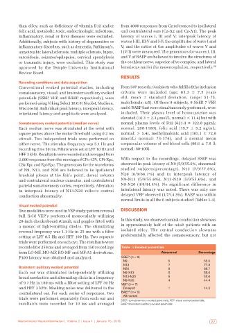
recorded for 250 ms and averaged from 150 recordings Table 1: Evoked potentials
from LO-MF, MO-MF, RO-MF and MF-A1 derivations. Abnormal Percentage
P100 latency was obtained and analyzed. SSEP (n = 9)
N9 5 55.6
N13 7 77.8
Brainstem auditory evoked potential N20 6 66.7
Each ear was stimulated independently utilizing N9‑N13 5 55.6
broad rarefaction and alternating clicks in a frequency N13‑N20 5 55.6
N9‑N20
of 9.7 Hz in 100 ms with a filter setting of LFF 30 Hz VEP (n = 7) 4 44.4
and HFF 3 kHz. Masking noise was delivered to the Delayed 1 14.3
contralateral ear. For each series of responses, two BAEP (n = 6)
All normal
trials were performed separately from each ear and SSEP: somatosensory evoked potentials; VEP: visual evoked potentials;
resultants were recorded for 10 ms and averaged BAEP: brainstem auditory evoked potentials
Neuroimmunol Neuroinflammation | Volume 2 | Issue 1 | January 15, 2015 27