
Page 30 - Read Online
P. 30
attacks, orthostatic hypotension, impaired heart-rate treatment with low doses of AChE-Is and/or steroids.
variability (HRV) [18,19] ). These significant cardiac The patients were admitted to the hospital for 24-48 h
manifestations were reported in patients with severe for the purpose of the research. Twenty healthy subjects
stages of MG and in the presence of thymoma. It has matched for age, sex and socioeconomic status were
been suggested that some MG patients may develop included in this study for statistical comparisons. Control
autonomic dysfunction and other nervous system subjects were recruited from the general population. This
manifestations. [18-26] It has also been suggested that the study was accepted by the regional Ethical Committee.
heart and skeletal muscle molecules are targets for the All patients and control subjects were briefed about the
autoimmune process of MG. [27-30] detailed information of this study and hence consented
to attend this study.
This study aimed to assess cardiac functions in
patients with MG using Ambulatory 24-h ECG Holter Excluded were subjects (patients and controls): (1)
Monitoring. HRV measures are sensitive indices with respiratory involvement or in crisis (i.e., severe
of cardiac autonomic function (sympathetic and stage of the disease); (2) with major systemic illness
parasympathetic). such as organic heart disease, diabetes, hypertension
or any other disease that might affect the autonomic
METHODS nervous system; (3) on medications known to affect
heart-rate/rhythm such as beta-blockers, vasopressors,
Subjects digitalis, theophylline, thyroid hormones, tricyclic
This study included 20 (males = 6, females = 14) antidepressant, antiarrythmic drugs, atropine and its
patients with MG without known cardiac diseases. Their derivatives, etc.; (4) with history of febrile illness in
age range is was 16-50 years and duration of illness the past 1-week; and (5) with lack of sound sleep the
ranged from 1 to 4 years. Clinical grading of the patients night prior to monitoring.
was done based on the medical-scientific advisory board
[4]
of the MG Foundation of America classification. Patients Measurements
grading was based on their histories and diagnosis shown All participants underwent conventional
in their medical records. Patients reported histories of echocardiography and Ambulatory ECG
weakness of ocular muscles (ptosis) (class I), with mild Holter-Monitoring. Holter monitoring is the continuous
and predominant weakness of limb muscles (class IIa) or 24-h monitoring of ECG activity of a patient’s heart
oropharyngeal muscles (class IIb), or with moderate and while engaged in daily activities. Ambulatory ECG
predominant weakness of limbs (class IIIa). Thymectomy was carried out using the 5-electrode Holter which
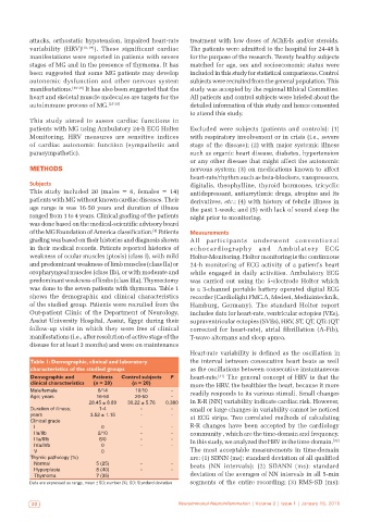
was done to the seven patients with thymoma. Table 1 is a 3-channel portable battery operated digital ECG
shows the demographic and clinical characteristics recorder (Cardiolight FMC.A, Medset, Medizintecknik,
of the studied group. Patients were recruited from the Hamburg, Germany). The standard Holter report
Out-patient Clinic of the Department of Neurology, includes data for heart-rate, ventricular ectopies (VEs),
Assiut University Hospital, Assiut, Egypt during their supraventricular ectopies (SVEs), HRV, ST, QT, QTc (QT
follow-up visits in which they were free of clinical corrected for heart-rate), atrial fibrillation (A-Fib),
manifestations (i.e., after resolution of active stage of the T-wave alternans and sleep apnea.
disease for at least 3 months) and were on maintenance
Heart-rate variability is defined as the oscillation in
Table 1: Demographic, clinical and laboratory the interval between consecutive heart beats as well
characteristics of the studied groups as the oscillations between consecutive instantaneous
Demographic and Patients Control subjects P heart-rate. [31] The general concept of HRV is that the
clinical characteristics (n = 20) (n = 20) more the HRV, the healthier the heart, because it more
Male/female 6/14 10/10 ‑ readily responds to its various stimuli. Small changes
Age; years 16‑50 20‑50 ‑
28.45 ± 8.89 30.22 ± 5.76 0.380 in R-R (NN) variability indicate cardiac risk. However,
Duration of illness; 1‑4 ‑ ‑ small or large changes in variability cannot be noticed
years 3.52 ± 1.15 ‑ ‑ at ECG strips. Two correlated methods of calculating
Clinical grade
I 0 ‑ ‑ R-R changes have been accepted by the cardiology
IIa/IIb 2/10 ‑ ‑ community , which are the time-domain and frequency.
IIIa/IIIb 8/0 ‑ ‑ In this study, we analyzed the HRV in the time-domain.
[32]
IVa/IVb 0 ‑ ‑
V 0 The most acceptable measurements in time-domain
Thymic pathology (%) are: (1) SDNN (ms): standard deviation of all qualified
Normal 5 (25) ‑ ‑ beats (NN intervals); (2) SDANN (ms): standard
Hyperplasia 8 (40) ‑ ‑
Thymoma 7 (35) deviation of the averages of NN intervals in all 5-min
Data are expressed as range, mean ± SD; number (%). SD: Standard deviation segments of the entire recording; (3) RMS-SD (ms):
22 Neuroimmunol Neuroinflammation | Volume 2 | Issue 1 | January 15, 2015