
Page 275 - Read Online
P. 275
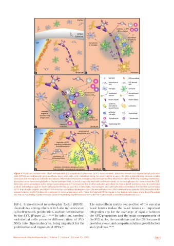
a                                 b


                         c


                         d                                              e
           Figure 3: Schematic representation of the demyelination and remyelination processes. (a) in a basal condition, type A neuroblasts and oligodendrocyte precursor
           cells (OPCs) are continuously generated from neural stem cells, with neuroblasts being the great majority progeny; (b) after a demyelinating episode, nearby
           astrocytes and microglia are activated and release inflammatory mediators, increasing the permeability of the blood‑brain barrier (BBB). By releasing chemokines,
           astrocytes recruit more microglial cells to the demyelinated area; (c) which phagocyte dead cells and myelin debris, as do macrophages that have crossed the BBB.
           Astrocytes and macrophages act as antigen-presenting cells to T lymphocytes that are then activated and attack the myelin sheath and dying cells. B lymphocytes
           produce autoantigens against myelin antigens functioning as opsonins; (d) microglia, macrophages, and astrocytes release mediators that mobilize parenchymal
           OPCs to proliferate, migrate, and differentiate into new myelinating oligodendrocytes in the demyelinated area. After a demyelinating episode, OPC production in the
           subventricular zone (SVZ) is favored in detriment of neuronal precursor cells. These SVZ-derived OPCs migrate to the demyelinated areas where they differentiate
           into mature myelinating oligodendrocytes; (e) new myelinating oligodendrocytes form a thinner myelin sheath around the demyelinated axon


           IGF‑1, brain‑derived neurotrophic factor  (BDNF),   The extracellular matrix composition of the vascular
           chemokines, among others, which also influence stem   basal lamina makes the basal lamina an important
           cell self‑renewal, proliferation, and fate determination   integration site for the exchange of signals between
           in the SVZ  [Figure  2]. [44‑46,49]  In addition, cerebral   the SVZ progenitors and the main compartments of
           endothelial cells  promote  differentiation  of  SVZ   the SVZ niche, the vasculature and the CSF, because it
           NSCs into oligodendrocytes, being important for the   provides, stores, and compartmentalizes growth factors
           proliferation and migration of OPCs. [42]          and cytokines. [42,45]


   266  Neuroimmunol Neuroinflammation | Volume 2 | Issue 4 | October 15, 2015  Neuroimmunol Neuroinflammation | Volume 2 | Issue 4 | October 15, 2015   267