
Page 274 - Read Online
P. 274
produce OPCs, which migrate radially out of the SVZ into oligodendrocytes, contributing to the remyelination
the surrounding cortex and white matter [Figure 2]. [5,38,39] process [Figure 3]. [4,5,41,42]
It should be noted that one NSC can generate either The generation of new oligodendrocytes from the
oligodendrocytes or neurons exclusively [13] and that SVZ is possible because adult NSCs, from which
the number of oligodendrocytes produced by the OPCs are derived, are embedded in the specialized
SVZ NSC pool is significantly inferior to the number and diverse microenvironments of the SVZ niche,
of olfactory interneurons. The relative quantity of which is responsible for regulating NSCs and
[5]
oligodendrocytes and neurons is area dependent: their progenies’ self‑renewal and differentiation
while in the posterior zone of the SVZ, the ratio is one by receiving information from the brain and other
oligodendrocyte to three neurons; in the rostral zone, tissues. [43‑45]
this ratio is 1:30. The ratio also changes dorsoventrally,
[5]
due to environmental cues. The dorsal part of the SVZ The first specialized microenvironment is the apical
is Wnt enriched, which favors OPC commitment. [13] ependymal compartment where slow‑dividing type B
On the other hand, the ventral part is more exposed cells are in direct contact with the cerebrospinal
to bone morphogenic proteins (BMP), which inhibits fluid (CSF) present in the space of the lateral
OPC specification. [40] ventricles, through a specialized apical process
surrounded by ependymal cells. [43,44,46] The adult
Contrary to the parenchymal OPCs, SVZ‑derived choroid plexus (CP) expresses and secretes to the
OPCs, although a minority in the brain, can CSF not only numerous trophic factors but also
migrate long distances into the corpus callosum, cytokines, which can influence the behavior of SVZ
striatum, and fimbria fornix, where they continue progenitor cells, modulating the self‑renewal capacity,
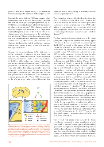
to divide or differentiate into mature myelinating proliferation, and differentiation [Figure 2]. [43,44]
and nonmyelinating oligodendrocytes. Adult SVZ Interleukin‑1β (IL‑1β) is one of the cytokines that
[5]
progenitor cells mostly generate neuronal lineage are secreted and regulated by CP, which acts on
cells and only a few oligodendrocytes; however, type B cells through binding to IL‑1 receptors to
progenitor cells show some lineage plasticity in up‑regulate vascular cell adhesion molecule 1
pathological conditions. In a demyelination context, expression, modulating type B cells adherence to
OPC production in SVZ is favored to the detriment of the SVZ niche. Insulin‑like growth factor 2 (IGF‑2)
neuronal precursor cells. These OPCs then migrate is also present in the adult CSF and regulates SVZ
to the affected areas, where they differentiate into progenitor proliferation. [44] Moreover, CP also secretes
guidance molecules, such as chemorepulsive factors
and chemoattractants, which regulate SVZ NSCs
and their progenies’ migration. [42] CP changes its
secretome under pathological conditions, leading to
a different CSF cellular and molecular composition
that will then influence the SVZ niche population.
For example, during peripheral tissue inflammation,
inflammatory information from the blood can have
an impact in the CNS. [47,48] Peripheral inflammation
elicits the up‑regulation of cytokines, adhesion
factors, and signaling pathway genes, such as tumor
necrosis factor‑α (TNF‑α), IL‑1β, and small inducible
cytokine A2 transcripts that are under the regulation
of the NF‑kB cascade, in the CP. [47] Then the CP,
through changes in CSF composition, will affect the
SVZ niche population.
The second SVZ niche component is the basal
vasculature, composed of blood vessels and a basal
Figure 2: Schematic representation of the adult subventricular zone (SVZ) lamina rich in laminin. [42] Here, type B cells have a long
neurogenic niche. SVZ lines the lateral ventricles and is comprised of three main specialized basal process through which they interact
cell types: the multipotent type B cells that give rise to type C cells (fast dividing
transient amplifying cells) that generate type A neuroblasts. Type B cells interact with blood vessels, and fast‑dividing/transit‑amplifying
basally with blood vessels and apically with the cerebrospinal fluid (CSF). The type C cells are also in very close proximity to blood
composition of the CSF is modified by the choroid plexus, a thin vascularized vessels. [43‑46] Endothelial cells secrete several diffusible
membrane mainly composed by epithelial cells, which secretes several cytokines
and trophic factors to the CSF signals, such as fibroblast growth factor‑2 (FGF2),
266 Neuroimmunol Neuroinflammation | Volume 2 | Issue 4 | October 15, 2015 Neuroimmunol Neuroinflammation | Volume 2 | Issue 4 | October 15, 2015 267