
Page 196 - Read Online
P. 196
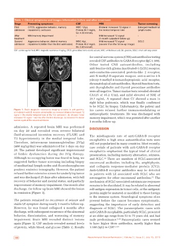
Table 1: Clinical symptoms and images information before and after treatment
           Stage    Presenting symptoms              CSF             MRI                       Tumor screening
           First    GTCS, aggressive behavior, memory   WBC 12/μL    Bilateral increased T2 signal in   Enlarged mediastinal
           admission  impairment, confusion          Protein 60.1 mg/dL  the medial temporal lobe  lymph nodes
                                                     Glc 4.30 mmol/L
           After    Mild memory impairment           ‑               Mild increased T2 signal   ‑
           treatment                                                 (1‑month outpatient follow‑up)
           Second   Aggressive behavior, distinct memory   WBC 4/μL  Mild increased T2 signal   SCLC
           admission  impairment (milder than the first admission)  Protein 40.2 mg/dL  (severer than the follow‑up image)
                                                     Glc 3.63 mmol/L
           CSF: cerebrospinal fluid, MRI: magnetic resonance imaging, GTCS: generalized tonic‑clonic seizure, WBC: white blood cell, Glc: glucose, SCLC: small cell lung cancer

                                                              for central nervous system (CNS) autoantibodies testing
                                                              revealed CSF antibodies to GABA-B receptor (IgG 1:100).
                                                              Other tested CNS autoantibodies, including
                                                              anti-leucine-rich glioma inactivated-1 (LGI1) receptor,
                                                              anti-contactin-associated protein-like 2 receptor,
                                                              anti-N-methyl-D-aspartate receptor, anti-a-amino-3-h
                    a              b                          ydroxy-5-methyl-4-isoxazolepropionic acid receptor,
                                                              rheumatological autoantibodies, thyroid function tests,
                                                              anti-thyroglobulin and thyroid peroxidase antibodies
                                                              were all negative. Tumor marker tests revealed elevated
                                                              CA125 of 35.2 U/mL and mild elevated of NSE of
                                                              25.7 ng/mL. A repeated chest CT showed a mass in
                                                              right hilus pulmonis, which was finally confirmed
                            c                                 to be SCLC by biopsy. Unfortunately, the patient and
           Figure  1: Brain magnetic resonance imaging screens in anti‑gamma‑  his  carers  refused  further  immunomodulating  or
           aminobutyric‑acid B receptor encephalitis: (a) revealed bilateral increased T2   antineoplastic treatments. He was discharged with
           signal in the medial temporal lobe at the first admission; (b) showed milder
           increased T2 signal 1 months after the first discharge; (c) revealed the lesion   memory impairment, which was persisted after another
           worsen again at the second admission
                                                              3 months follow-up.
           admission.  A  repeated  brain  MRI  was  performed   DISCUSSION
           on day 20 and revealed even severer bilateral
           fluid-attenuated  inversion  recovery  (FLAIR)  and   The misdiagnosis  rate of anti-GABA-B receptor
           T2  hyperintensity  in  the  medial  temporal  lobe.   encephalitis is high since autoantibodies tests were
           Therefore, intravenous immunoglobulins  (IVIg)     still not popularized in many countries. Most recently,
           (400 mg/kg/day) was administered for 5 days on day   case  serials  of  patients  with  anti-GABA-B  receptor
           20. The patient developed significant improvement   encephalitis emphasized the typical triad of clinical
           of limbic dysfunction during the IVIg therapy.     presentation, including memory alternation, seizures,
           Although no occupying lesion was found in lung, we   and SCLC.  There are numbers of SCLC-associated
                                                                        [6]
           suggested further tumor screening including biopsy   onconeural antibodies, including Hu, amphiphysin,
           of mediastinal lymph nodes and fluorodeoxyglucose   and collapsin response-mediator protein  (CV2).
           positron emission tomography. However, the patient   Anti-GABA-B receptor antibodies may be detected
           refused further extensive screen for underlying tumor   in patients with LE associated with SCLC who are
           and was discharged 25 days after admission, with full   seronegative for other onconeural antibodies.  The
                                                                                                         [7]
           recovery of behavior and mental status, and partially   mechanism of SCLC-associated autoimmune responses
           improvement of memory impairment. One month after   remains to be elucidated. It may be related to abnormal
           discharge, the follow-up brain MRI showed the lesion   self-antigen expression in tumor cells, or the antigenic
           in remission [Figure 1].                           protein might be mutated or modified to those foreign
                                                              to the immune system. Neurological symptoms often
           The patients remained no recurrence of seizure and   present  before  the  cancer  becomes  symptomatic,
           stable LE symptom during nearly 3 months follow-up.   suggesting the importance of early detection and
           However,  he  was  found  collapsed  4  months  after   diagnosis of SCLC. The paraneoplastic cases with
           discharge, with subsequent recurrence of the abnormal   anti-GABA-B encephalitis, particularly SCLC, presented
           behavior, disorientation, and worsening of memory   at an older age range from 53 to 75 years old, and had
           impairment. Brain MRI revealed distinct lesions    male predominance. [1,4]  Paraneoplastic cases seemed
           again [Figure 1]. CSF analysis revealed normal levels   to have higher titers antibodies, mostly higher than
           of protein, white blood, and glucose [Table 1]. Results   1:100 (IgG) in CSF. [1,4,8]


            188                                              Neuroimmunol Neuroinflammation | Volume 2 | Issue 3 | July 15, 2015  Neuroimmunol Neuroinflammation | Volume 2 | Issue 3 | July 15, 2015                              189