
Page 192 - Read Online
P. 192
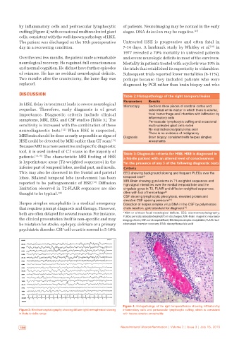
by inflammatory cells and perivascular lymphocytic   of patients. Neuroimaging may be normal in the early
           cuffing [Figure 4] with occasional multinucleated giant   stages. DNA detection may be negative. [17]
           cells, consistent with the well-known pathology of HSE.
           The patient was discharged on the 10th postoperative   Untreated HSE  is progressive  and  often fatal  in
           day in a recovering condition.                     7-14 days. A landmark study by Whitley et al. [18]  in
                                                              1977 revealed a 70% mortality in untreated patients
           Over the next few months, the patient made a remarkable   and severe neurologic deficits in most of the survivors.
           neurological recovery. He regained full consciousness   Mortality in patients treated with acyclovir was 19% in
           and normal cognition. He did not have further episodes   the trials that established its superiority to vidarabine.
           of seizures. He has no residual neurological deficits.   Subsequent trials reported lower mortalities (6-11%),
           Two months after the craniectomy, the bone flap was   perhaps because they included patients who were
           replaced.                                          diagnosed by PCR rather than brain biopsy and who

           DISCUSSION
                                                               Table 2: Histopathology of the right temporal lesion
                                                               Parameters  Results
           In HSE, delay in treatment leads to severe neurological   Microscopy  Sections show pieces of cerebral cortex and
           sequelae. Therefore, early diagnosis is of great                subcortical white matter in which there is edema,
           importance. Diagnostic criteria include clinical                focal hemorrhage and infarction with infiltration by
           symptoms, MRI, EEG, and CSF studies [Table 3]. The              inflammatory cells
                                                                           Perivascular lymphocytic cuffing and occasional
           sensitivity is increased with the combination of these          multinucleated giant cells noted
           neurodiagnostic tests. [12]  When HSE is suspected,             No viral inclusions/granuloma seen
           MRI brain should be done as early as possible as signs of   Diagnosis  There is no evidence of malignancy
                                                                           Brain biopsy: consistent with herpes simplex
           HSE could be detected by MRI earlier than CT scan. [13]         encephalitis
           Because MRI is a more sensitive and specific diagnostic
           tool, it is used instead of CT scans in the majority of   Table 3: Diagnostic criteria for HSE. HSE is diagnosed in
           patients. [13-15]  The characteristic MRI finding of HSE   a febrile patient with an altered level of consciousness
           is hyperintense areas (T2-weighted sequences) in the   *in the presence of any 3 of the following diagnostic tests
           inferior part of temporal lobes, medial part, and insula.   Criteria
           This may also be observed in the frontal and parietal   EEG showing background slowing and frequent PLEDs over the
           lobes. Bilateral temporal lobe involvement has been   temporal lobe [8]
           reported to be pathognomonic of HSE. [16]  Diffusion   MRI‑Brain showing gyral edema in T1‑weighted sequences and
                                                               high signal intensities over the medial temporal lobe and the
           limitation observed in T2-FLAIR sequences are also   cingulate gyrus in T2, FLAIR and diffusion‑weighted sequences,
           thought to be typical. [16]                         often with foci of hemorrhage [9]
                                                               CSF showing lymphocytic pleocytosis, elevated protein and
                                                               elevated CSF opening pressure [10]
           Herpes simplex encephalitis is a medical emergency   Detection of herpes simplex virus DNA in the CSF by polymerase
           that requires prompt diagnosis and therapy. However,   chain reaction: gold standard for diagnosis [11]
           both are often delayed for several reasons. For instance,   *With or without focal neurological deficits. EEG: electroencephalography;
           the clinical presentation itself is non-specific and may   PLEDs: periodic lateralized epileptiform discharges; MRI‑Brain: magnetic resonance
                                                              imaging of brain; CSF: cerebrospinal fluid; HSE: herpes simplex encephalitis; FLAIR: fluid‑
           be mistaken for stroke, epilepsy, delirium or a primary   attenuated inversion recovery; DNA: deoxyribonucleic acid
           psychiatric disorder. CSF cell count is normal in 5-10%


                                                              Figure 4: Histopathology of the right temporal lesion showing infiltration by
           Figure 3: Electroencephalography showing diffuse right hemispherical slowing   inflammatory cells and perivascular lymphocytic cuffing, which is consistent
           in theta to delta range                            with herpes simplex encephalitis


            184                                              Neuroimmunol Neuroinflammation | Volume 2 | Issue 3 | July 15, 2015  Neuroimmunol Neuroinflammation | Volume 2 | Issue 3 | July 15, 2015                              185