
Page 193 - Read Online
P. 193
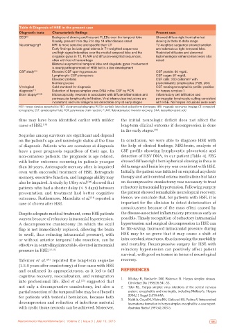
Table 4: Diagnosis of HSE in the present case
           Diagnostic tests  Characteristic findings                            Present case
           EEG [8]         Background slowing and frequent PLEDs over the temporal lobe  Showed diffuse right hemispherical
                           Usually, present from day 2 to day 14 after disease onset  slowing in theta to delta range
           Neuroimaging [9]  MRI is more sensitive and specific than CT         T2‑weighted sequence showed swollen
                           Early findings include gyral edema in T1‑weighted sequences   and edematous right temporal lobe.
                           and high signal intensities over the medial temporal lobe and the   Restricted diffusion and abnormal
                           cingulate gyrus in T2, FLAIR and diffusion‑weighted sequences,   leptomeningeal enhancement were also
                           often with foci of hemorrhage                        noted
                           Bilateral assymetrical temporal lobe and cingulate gyrus involvement
                           is nearly pathognomonic of HSE but is a late development
           CSF study [10]  Elevated CSF opening pressure                        CSF protein: 60 mg/dL
                           Lymphocytic CSF pleocytosis                          CSF sugar: 87 mg/dL
                                                                                                 3
                           Elevated proteins                                    CSF cells: 200 cells/mm  with
                           Normal glucose                                       predominantly lymphocytes (P28, L64)
           Virological     Gold standard for diagnosis                          CSF meningoencephalitis profile: positive
           diagnosis [11]  Detection of herpes simplex virus DNA in the CSF by PCR  for herpes simplex‑1
           Brain biopsy [27,28]  Microscopically, necrosis is associated with diffuse inflammation and   Inflammatory cell infiltration and
                           perivascular lymphocytic infiltration. Viral intranuclear inclusions are   perivascular lymphocytic cuffing consistent
                           inconstant, and viral antigens are detectable only at early stages  with HSE. No herpes inclusions were seen
           HSE: herpes simplex encephalitis; EEG: electroencephalography; PLEDs: periodic lateralized epileptiform discharges; MRI: magnetic resonance imaging; CT: computed
           tomography; CSF: cerebrospinal fluid; PCR: polymerase chain reaction; FLAIR: fluid‑attenuated inversion recovery; DNA: deoxyribonucleic acid

           thus may have been identified earlier with milder   the initial neurologic deficit does not affect the
           cases of HSE. [19]                                 long-term clinical outcome if decompression is done
                                                              in the early stages. [26]
           Sequelae among survivors are significant and depend
           on the patient’s age and neurologic status at the time   In conclusion, we were able to diagnose HSE with
           of diagnosis. Patients who are comatose at diagnosis   the help of clinical findings, MRI-brain, analysis of
           have  a  poor  prognosis  regardless  of  their  age.  In   CSF profile showing lymphocytic pleocytosis and
           non-comatose patients, the prognosis is age related,   detection of HSV DNA, in our patient [Table 4]. EEG
           with better outcomes occurring in patients younger   showed diffuse right hemispherical slowing in theta to
           than 30 years. Anterograde memory often is impaired   delta range and brain biopsy was consistent with HSE.
           even with successful treatment of HSE. Retrograde   Initially, the patient was initiated on empirical acyclovir
           memory, executive function, and language ability may   therapy and anti-cerebral edema medications but later
           also be impaired. A study by Utley et al. [20]  showed that   on decompressive craniectomy was necessary to treat
           patients who had a shorter delay (< 5 days) between   refractory intracranial hypertension. Following surgery
           presentation and treatment had better cognitive    the patient showed remarkable neurological recovery.
           outcomes. Furthermore, Marschitz et al. [21]  reported a   Hence, we conclude that, for patients with HSE, it is
           case of chorea after HSE.                          important for the clinician to detect deterioration of
                                                              consciousness because of the mass effect caused by
           Despite adequate medical treatment, some HSE patients   the disease-associated inflammatory process as early as
           worsen because of refractory intracranial hypertension.   possible. Timely recognition of refractory intracranial
           A  decompressive craniectomy  (in which the skull   hypertension and surgical decompression in HSE can
           flap is not immediately replaced, allowing the brain   be life-saving. Increased intracranial pressure during
           to swell, thus reducing intracranial pressure), with   HSE may be so grave that it may cause a shift of
           or without anterior temporal lobe resection, can be   intra-cerebral structures, thus increasing the morbidity
           effective in controlling intractable, elevated intracranial   and mortality. Decompressive surgery for HSE with
           pressure in HSE. [22,23]                           refractory hypertension can positively affect patient
                                                              survival, with good outcomes in terms of neurological
           Taferner  et  al. [24]  reported the long-term sequelae   recovery.
           (1.5-8 years after craniectomy) of four cases with HSE
           and confirmed its appropriateness, as it led to full   REFERENCES
           cognitive recovery, resocialization, and reintegration
           into professional life. Ebel  et  al. [25]  suggested that   1.   Whitley R, Kimberlin DW, Roizman B. Herpes simplex viruses.
                                                                  Clin Infect Dis 1998;26:541‑53.
           not only a decompressive craniectomy, but also a   2.   Tyler KL. Herpes simplex virus infections of the central nervous
           partial resection of the temporal lobe may be of benefit   system: encephalitis and meningitis, including Mollaret’s. Herpes
           for patients with tentorial herniation, because both   2004;11 Suppl 2:57A‑64A.
           decompression and reduction of infectious material   3.   Malik A, Goyal M, Mishra NK, Gaikwad SB, Padma V. Intracerebral
                                                                  haematoma formation in herpes simplex encephalitis: a case report.
           with cystic tissue necrosis can be achieved. Moreover,   Australas Radiol 1997;41:303‑5.


   184  Neuroimmunol Neuroinflammation | Volume 2 | Issue 3 | July 15, 2015  Neuroimmunol Neuroinflammation | Volume 2 | Issue 3 | July 15, 2015   185