
Page 18 - Read Online
P. 18
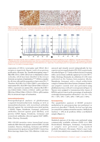
Figure 1: Exemplary images of immunohistochemical evaluation of isocitrate dehydrogenase (IDH‑1 [R132H]) and karyopherin a2 (KPNA2) expression as well as
           nuclear accumulation of p53 protein. Immunohistochemical staining with antibodies against mutated IDH‑1 (R132H) shows a strong cytoplasmic immunoreactivity
           in the tumor cells. Nuclear expression of KPNA2 is detected in a subpopulation of tumor cells. Nuclear p53 accumulation can be observed in > 50% of the tumor


                         a                                    b
           Figure 2: Pyrosequencing was used for quantitative analysis of O ‑methylguanine DNA methyltransferase promoter methylation. Pyrogram demonstrating (a) an
                                                    6
           unmethylated and (b) methylated glioblastoma tissue. Each colored box includes one of the four studied CpG positions (CpGs 9‑12). The incorporation of the bases
           guanine and adenine represent the methylated and unmethlylated fractions, respectively. The percentages given in both pyrograms reflect the methylated fractions
           (fractions over 10% define a methylated sample)
           expression of CD11c  (a-integrin) and CD123 (IL-3   assessed and visually scored independently by two
           receptor a), respectively. Figures 1 and 2 of our previous   experienced neuropathologists (PN, GHG) as previously
           publication illustrate the classification/gating steps.    described [Figure 1]. [14]  Immunohistochemical staining
                                                         [4]
           HLA-DR, CD11c, CD45, CD123 are co-stimulatory surface   with a monoclonal antibody against p53 (clone DO-7,
           molecules, which have been identified in the relevant   Dako, Glostrup, Denmark), in a dilution of 1:150, were
           literature as markers of maturation. [24-27]  CD34 is a marker   performed on the Ventana Immunostainer  (Roche,
           for all stem cells and the proportion of precursor DCs. [28]    Mannheim,  Germany),  with  a  closed  avidin-biotin
           Therefore, HLA DR+/Lin-/CD34-/CD45+ were defined as   complex Ventana Detection System (Ventana). Positive
           total mature DCs. HLA DR+/Lin-/CD34-/CD45+/CD123-/  and negative controls were also performed using
           CD11c- represent less mature DCs, whereas HLA DR+/  glioblastoma tissue with p53 overexpression [Figure 1].
           Lin-/CD34-/CD45+/CD11c+/CD123- mDCs and HLA        Tumors were assigned to immunoreactivity classes of
           DR+/Lin-/CD34-/CD45+/CD123+/CD11c- pDCs mature     KPNA2 and p53 based on the percentage of moderately
           DCs in advanced stages of maturation.              or strongly immunopositive cell nuclei (< 1%, 1%-< 5%,
                                                              5%‑< 10%, 10%‑< 20%, 20%‑< 50% and ≥ 50%).
           Immunohistochemistry
           Neuropathological analysis of  glioblastomas       Pyrosequencing
           comprised hematoxylin/eosin staining as well as    The quantitative analysis of MGMT promoter
           immunohistochemistry with monoclonal antibodies    methylation by pyrosequencing was performed as
           directed against the microtubule-associated protein   previously described. [29]  Briefly, the first four CpG sites
           2 (MAP2, Sigma, Steinheim, Germany), polyclonal    are assayed for a primer extension reaction. Methylated
           antibodies  directed  against  glial  fibrillary  acid   fractions > 10% at all positions define a methylated
           protein  (GFAP, Sigma, Steinheim, Germany) and     sample [Figure 2].
           monoclonal antibodies directed against Ki67 (MIB1,
           Dako, Glastrop, Denmark).                          Statistical analysis
                                                              Statistical analyses of the data were performed using
           IDH-1-R132H mutation status  (monoclonal mouse     commercially available  software  (SSPS 21.0,  IBM
           antibody H09 directed against mutated IDH-1 R132H   Deutschland, Ehningen, Germany). Comparisons
           mutation, Dianova, Hamburg, Germany) and KPNA2     of samples were performed using standard
           immunoreactivity (goat polyclonal SC6917; Santa Cruz   methods (Pearson’s Chi-square, Fisher’s exact test).
           Biotechnology, Santa Cruz, USA; dilution 1:100) were   P < 0.05 (two-tailed) were considered to be statistically


            10                                             Neuroimmunol Neuroinflammation | Volume 2 | Issue 1 | January 15, 2015